International Journal of Chemical Engineering and Analytical Science, Vol. 1, No. 2, November 2016 Publish Date: Aug. 16, 2016 Pages: 77-83
Synthesis, Characterization and Anti-prostatic Hyperplasia Activity of Zn(II)-1-(5-Hydroxy-3-methyl-1-phenyl-1-pyrazol-4-yl)octadecan-1-one Complex
C. E. Sokwaibe, I. E. Otuokere*
Department of Chemistry, Michael Okpara University of Agriculture, Umudike, Nigeria
Abstract
Zinc(II)1-(5-Hydroxy-3-methyl-1-phenyl-1-pyrazol-4-yl)octadecan-1-one complex was synthesized by the reaction of 1-(5-Hydroxy-3-methyl-1-phenyl-1-pyrazol-4-yl)octadecan-1-one with zinc(II)sulphate heptahydrate. A dirty-white precipitate with a melting point of 310°C was isolated. The complex was insoluble in water but soluble in organic solvent. The molar conductance value of the complex was 4.8 Siemens mol-1cm-1 which suggested that the complex is covalent. Infrared, 1HNMR and 13CNMR characterization showed that the complex is tetrahedral. The results of the anti-prostatic hyperplasia studies showed a significant increase (P<0.05) in Prostate specific antigen (PSA) value of hormone control group (HC) receiving only hormonal induction relative to [Zn.HMPPO.H2O] group. Serum prostatic acid phosphatase activity (PACP) showed a significant increase (P<0.05) in ‘HC’ Group receiving only hormonal induction relative to [Zn.HMPPO.H2O] group. Relative prostate weight was significantly increased (P<0.05) in ‘HC’ group compared to [Zn.HMPPO.H2O] group. We deduced that zinc (II)1-(5-Hydroxy-3-methyl-1-phenyl-1-pyrazol-4-yl)octadecan-1-one inhibited the growth of benign prostatic hyperplasia.
Keywords
Zinc(II)1-(5-Hydroxy-3-methyl-1-phenyl-1-pyrazol-4-yl)octadecan-1-one, Benign Prostatic Hyperplasia, Complex, Characterization
Received: July 8, 2016
Accepted: July 25, 2016
Published online: August 16, 2016
@ 2016 The Authors. Published by American Institute of Science. This Open Access article is under the CC BY license. http://creativecommons.org/licenses/by/4.0/
Contents
1. Introduction 2. Methodology 2.1. Chemical and Solvents 2.2. Physical Measurements 2.3. Synthesis of Zinc(II) 1-(5-hydroxy-3-methyl-1-phenyl-1H-pyrazol-4-yl)octadecan-1-one 2.4. Biological Activity 2.5. Preparation of the Hormone 2.6. Experimental Design 2.7. Sample Collection and Analysis 3. Results 4. Discussion 5. Conclusion
1. Introduction
Coordination compounds consist of a central metal ion surrounded by a suitable number of ligands in a demonstrated geometrical arrangement. The major breakthrough in this field was in 1893 when Alfred Werner synthesized cobalt complexes containing chlorine and ammonia. He won a noble prize for his contribution to knowledge in 1913. The role of transition metal complexes in the field of inorganic chemistry and medicinal chemistry is becoming increasingly important. Recent advances in inorganic chemistry have made possible formation of number of transition metal complexes with organic ligand of interest, which can be used as therapeutic agent. The use of transition metal complexes as therapeutic compounds has become more and more pronounced.
Neurological disorders have been successfully managed with transition metal complexes [1]. Complexes of gold nanoparticles enhance DNA damage and make the treatment target specific when used in combination with radio therapy or chemotherapy [2]. Various forms of cancer have been treated with Lanthanum [3]. Mn (III) complexes have been reported by Ansari and colleagues to induce tumor selective apoptosis of human cells [4]. Improved glycemia among patients with diabetes have been significantly improved with chromium supplementation but do not show any significant effect on glucose metabolism in healthy individuals [5]. Remarkable anti-proliferative properties of some iron chelates have been reported [6-8]. In 2008, the anti-cancer activity of gold complexes was reported [9]. The mechanism of action of gold complexes was different form cisplain because it attacked the mitochondria of the cancer cells and not the DNA [9]. Silver complex of chlorohexidine- sulfadiazine is effective against catheter infections in vivo [10]. Trypanocidal activity of Pt, Rh, Ir, Pd, and Os organometallics were reported by Lorisean and colleagues in 1992 [11].
The role of zinc in maintenance hemodialysis patients were studied by Fukushima and colleagues [12]. Few attempts have been made in search of applications for zinc complexes. Zinc complexes containing sulphur as donor atoms has been reported to exhibit strong affinity to lipid-rich regions and possibly the treatment of ischemic heart disease [13]. Zincgluconate lozenges has been given favorable attention for the relief of common cold symptoms [14,15]. Endocrinological, neurological and immunological activity of zinc complexes have been reported in recent years [16,17]. The effect of zinc in the management of Alzheimer’s disease is quite interesting but still ambiguous [18]. Intake of higher zinc has also been associated with a slightly lower risk of type 2 diabetes in women [8]. Mice infected with plasmodium berghei were administered 60 mg/kg of o-vanillin- (4-methylthiosemicarbazone) and o-vanillin- (4-phenylthiosemicarbazone) complexes. These complexes exhibit antimalarial activity in the infected mice [19].
In continuation with the current research on the applicability of zinc complexes, we report the synthesis, characterization and anti-prostatic hyperplasia activity of Zn(II) complex of 1-(5-hydroxy-3-methyl-1-phenyl-1-pyrazol-4-yl) octadecan-1-one
2. Methodology
2.1. Chemical and Solvents
The chemicals and solvents used in this study were of analytical grade and used as obtained from sigma-Aldrich chemical company without further purification. The chemicals are; β-estradiol-17-valerate and 5α-androstan-17β-ol-3-one (hormone), zinc(II)sulphateheptahydrate, 3-methyl-1-phenylpyrazol-5-one, octadecanoyl chloride, calcium hydroxide.
2.2. Physical Measurements
The melting point of the complex was determined using Gallenkamp melting point apparatus. The molar conductance of the complex was determined in either DMF or DMSO using JenwaypeMJ conductivity meter with a cell constant of 1.05. Atomic absorption spectroscopy and elemental analysis were carried out on Varian AA spectrometer (AA240FS). 0.2 g of complex was digested with 2.0cm3 of 50% of water/nitric acid. The digest was rinsed quantitatively into a 100 ml standard flask and made up to the mark with deionized water. The elemental analysis for C,H and N were obtained using a Perkin-Elmer 240B elemental analysis instrument. Infrared spectrum was collected on Perkin Elmer Paragon 1000 FT-IR spectrophotometer equipped with caesium iodide (4000-250 cm-1) as KBr pellets. The Proton and Carbon-13 NMR spectra were obtained on a BrukerAvance III HD spectrometer operated at a frequency of 600 MHz in Laboratory F22 of the Department of Chemistry, Rhodes University, South Africa. Dimethylsulfoxide (DMSO) was used as the solvent.
2.3. Synthesis of Zinc(II) 1-(5-hydroxy-3-methyl-1-phenyl-1H-pyrazol-4-yl)octadecan-1-one
1-(5-hydroxy-3-methyl-1-phenyl-1H-pyrazol-4-yl) octadecan-1-one (HMPPO ligand) was synthesized as described in previous publication [20]. The complex was prepared following reported procedure [21,22]. Zn (II) solutions were prepared by dissolving 5.94g (0.025mole) ZnCl2.7H2O, in 100ml of deionized water with warming at 90°C. The solution was added to 22g (0.025mole) of HMPPO ligand (complexing agent) in 100ml ethanol solution at 70°C. The complex was allowed to cool and crystals that appeared was washed, recrystalized from aqueous ethanol (1:1), filtered and dried in a desiccator.
2.4. Biological Activity
Animal Housing
A total of 15 male albino rats having an average weight of about 104.12g each were purchased from an animal breeding unit in the department of veterinary Pharmacology, University of Nigeria. The rats were housed in standard steel cages with a plastic base and acclimatized for 7 days under humid tropical condition in the animal house of the college of Natural and Applied Science, Department of Biochemistry, Michael Okpara University of Agriculture, Umudike, Abia State. The rats were exposed to 12hr light/dark cycle and were given free access to clean tap water and commercial rat chow purchased from vital feeds Nigeria limited.
2.5. Preparation of the Hormone
This was prepared following outlined literature [23]. 250mg dihydrotestosterone and 25mg estradiolvalerate were dissolved in 25ml olive oil and administered intravenously. The dose of the complex [Zn.HMPPO.H2O] was formulated as follows; 0.125g of the complex was dissolved in 25ml of olive oil to yield a stock of 5mg/ml.
2.6. Experimental Design
After 7 days of acclimatization, the experimental animals were randomly assigned into three (3) experimental groups of 3 rats each. The groups were labeled as follows.
Group 1 – Standard test control group (STC group): Received no hormone treatment and no [Zn.HMPPO.H2O] administration but was fed normal diet for 28 days. This group served as the standard control group.
Group 2 – [Zn.HMPPO.H2O] group: Received subcutaneous injection of the hormone every day and 5mg/ml oral administration of [Zn.(HMPPO).H2O] everyday for 28days.
Group 3 – Hormone control group (HC group): Received subcutaneous injection of the hormone every day with no oral administration of [Zn.(HMPPO).H2O] for 28 days. This group served as hormone control group
2.7. Sample Collection and Analysis
After 28 days, all the rats were weighed before they were sacrificed by cardiac puncture after dazing with a cervical blow. They were bled exhaustively. Serum collection was done according to method reported in literature [24]. Whole blood was collected in a vacutainer. The blood was allowed to clot by leaving it undisturbed at room temperature for 20 minutes. The clot was removed by centrifuging at 2,000 rpm for 5 minutes in a refrigerated centrifuge. The liquid component (serum) was transferred into a clean polypropylene tube using a Pasteur pipette. Vital organs for the study were excised and weighed. Prostate Specific Antigen (PSA) and Prostate Acid Phosphate (PACP) analysis were done following the procedure outlined in literature [23]. Relative organ weight determination was calculated as the ratio of the excised organ and the final weight of each rat and the mean taken for each group. Statistical analysis results were expressed as simple mean and standard deviation, mean were analyzed for difference using ANOVA, SPSS 17 software.
3. Results
The physical and analytical data, solubility, infrared, 1HNMR and 13CNMR are presented in Tables 1 – 5 respectively. Graph of PSA concentration in rats, PACP concentration graph in rats and mean prostatic weight graph in rats are presented in Figures 1 – 3 respectively.
Table 1. Physical and microanalytical data for zinc complex.

Table 2. Solubility data zinc complex in various solvents.
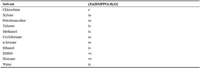
Legend: s = soluble, is = insoluble, ss = slightly soluble, vs = very soluble.
Table 3. Selected IR bands for the [Zn(HMPPO).H2O] complex.
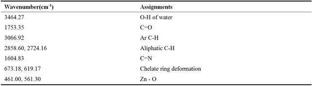
Table 4. 10 1H NMR Bands of [Zn(HMPPO).H2O] (Complex).
![]()
Table 5. 13C NMR Bands of [Zn(HMPPO).H2O] (Complex).
![]()
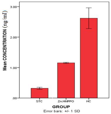
Figure 1. Graph of PSA concentration in rats.
Values are significantly different (p>0.05) from one another
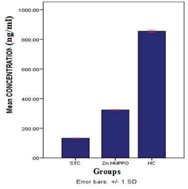
Figure 2. PACP concentration graph in rats.
Values are significantly different (p>0.05) from one another
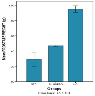
Figure 3. Mean prostatic weight graph in rats.
Values are significantly different (p>0.05) from one another
4. Discussion
The formation of a dirty-white precipitate showed the formation of zinc complex because zinc is a d10 metal. The mole ratio of the ligand and metal was confirmed to be 1:1 based on the elemental analysis data. The calculated and experimental elemental analyses are in good agreement. The conductivity measurement of the complex in acetone (10-3 M) was 4.8 Siemens mol-1cm-1. This value is lower than 30 Siemens mol-1cm-1, showing that it is a non-ionic compound [25,26]. Based on the solubility data, the complex was proposed to be hydrophobic. The complex was insoluble in water. The complex showed high degrees of solubility in ethylacetate, acetone, DMSO, THF, dioxane and pyridine. These solvents have lone pairs of electrons, which they must have donated to the complexes to complete the octahedron in the complexes, thereby reducing further the ionic character of the complex, thus an increase in the solubility of the complexes in those solvents.
The vibration frequency 3464.27 cm-1 was assigned OH of coordinated water in the complex (Table 3). This band is absent in the infrared of the free ligand [20]. The νC=O stretching bands of HMPPO was observed as a very strong peak at 1743 cm-1 [20] but on coordination with zinc it shifted to 1753.35cm-1. This observation indicates the involvement of the C=O group in the chelation process, hence the formation of C=O—>M bonding system. C=N vibration frequency band of HMPPO was observed as strong peak at 1618.33 cm-1 [20], but in the complex it shifted to1604.83cm-1, this indicates the involvement of the C=N in bonding system. Aromatic C-H band was observed at 3066.92 cm-1 while the aliphatic C-H bands were observed at 2858.6 and 2724.16 cm-1. Chelate ring deformations were observed 673.18 and 619.17 cm-1. The vibration frequency 461.00 and 561.30 cm-1 in the zinc complex was assigned Zn – O bond [22]. These bands were absent in the free ligand [20].
The signals of methyl and methylene protons were observed as multiplets at 1.17 – 1.59 ppm, while the phenyl protons were observed at chemical shifts 7.34 – 7.95 ppm (Table 4). 13C NMR spectral data (Table 5) indicated that the carbon resonance spectra of C=O was observed at chemical shift value of 188.40 ppm. This value indicate C=O in complexation with zinc because of the vibrational shift as compared to the free ligand in our previous publication [20]. The aromatic carbons were observed at 111.53 – 164.42 ppm. The carbon atoms of methyl and methylene were observed at 14.43 – 19.36 ppm.
Based on the elemental analyses, infrared spectrum and NMR characterization, the proposed structure of the complex is presented in Figure 4
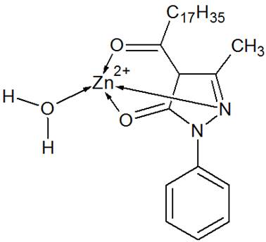
Figure 4. Proposed structure of zinc(II) 1-(5-Hydroxy-3-methyl-1-phenyl-1-pyrazol-4-yl)octadecan-1-one complex.
Benign Prostatic Hyperplasia results from the enlargement of the prostate gland due to proliferation of prostate stoma and epithelium in the transitional zone [27]. This proliferation cause lower urinary tract symptoms (LUTS) such as difficulty in urination or decrease in maximum flow rate, increase in residual urine volume in the bladder and increase in prostate size [28]. PSA is believed to be synthesized in the rough endoplasmic reticulum, stored in the vesicle and vacuole and released in the glandular Lumina by exocytosis [29]. Because of its tissue specificity, PSA is used as a maker for BPH and prostate cancer. PSA is a single polypeptide and occurs both in normal and malignant prostatic tissues and in the gland of men with BPH, but not in any other human tissue [29]. PSA is a secretory product of prostatic cells and will increase when the prostatic cells increase in number. PSA value can be used to predict enlargement of the prostate because high PSA value correlate increase in prostate mass [28]. Prostatic Acid Phosphates (PACP) could be used as a marker to detect prostate disorders in human or animals [30]. However, the discovery of PSA has resulted in a shift from PACP to PSA. Elevated PACP level in animals treated with DHT and estradiol have been reported [31] and this may be due to hyperplasia of the prostate gland. Together with PSA, PACP can give useful information about prostatic disease especially BPH prostate cancer
The result shows a significant increase (P<0.05) in PSA value of ‘HC’ group receiving only hormonal induction relative to [Zn.HMPPO.H2O] group. Serum prostatic acid phosphatase activity showed a significant increase (P<0.05) in ‘HC’ Group receiving only hormonal induction relative to [Zn.HMPPO.H2O] group. Relative prostate weight was significantly increased (P<0.05) in ‘HC’ group compared to [Zn.HMPPO.H2O] group. The development of antitumor drugs based on transition metal complexes is currently a very active field [32–34].
5. Conclusion
Infrared, 1HNMR and 13CNMR spectroscopic characterization showed that the complex is tetrahedral. 1-(5-Hydroxy-3-methyl-1-phenyl-1-pyrazol-4-yl)octadecan-1-one coordinated to zinc through C=O, OH and C=N. The complex is a dirty-white covalent hydrophobic solid with a melting point of 310°C. Our studies showed that the complex inhibited benign prostatic hyperplasia. We recommend the investigation of the toxicity of the complex.
References
- Hashimoto, R., Fujimaki, K., Jeong, M.R., Senatorov, V.V., Christ, L., Leeds,P.,Chuang,D.M.,Takeda, M. (2003). Neuroprotective actions of lithium.SeishinShinkeigakuZasshi, 105(1): 81-86.
- Zheng, Y., Sanche, L. (2009). Gold nanoparticles enhance DNA damage induced by anti-cancer drugs and radiation. Radiat. Res. 172(1): 114-119.
- Kapoor, S. (2009). Lanthanum and its rapidly emerging role as an anticarcinogenic agent.J. Cell Biochem.106(2): 193.
- Ansari, K.I., Grant, J.D., Kasiri, S., Woldemariam, G., Shrestha, B., Mandal, S.S. (2009). Manganese (III)-salens induce tumor selective apoptosis inhuman cells. J. Inorg. Biochem. 103(5): 818-826.
- Balk, E.M., Tatsioni, A., Lichtenstein, A.H., Lau, J., Pittas, A.G. (2007). Effect of chromium supplementation on glucose metabolism and lipids.A systematic review of randomized controlled trials.Diabetes Care. 30(8): 2154-2163.
- Lange, T.S., Kim, K.K., Singh, R.K., Strongin, R.M., McCourt, C.K., Brard, L. (2008). Iron (III)-salophene: an organometallic compound with selective cytotoxic and anti-proliferative properties in platinum-resistant ovarian cancer cells. PLoS One. 3(5): e2303.
- Ray, S., Mohan, R., Singh, J.K., Samantaray, M.K., Shaikh, M.M., Panda, D.,Ghosh, P. (2007). Anticancer and antimicrobial metallopharmaceutical agents based on palladium, gold, and silver N-heterocyclic carbine complexes. J. Am. Chem. Soc. 129(48): 15042-15053.
- Sun, Q. vanDam, R.M., Willett, W.C., Hu, F.B. (2009). Prospective study of zinc intake and risk of type 2 diabetes in women.Diabetes Care. 32(4): 629-634.
- Au, L., Zheng, D., Zhou, F., Li, Z.Y., Li, X., Xia, Y. (2008). A quantitative study on the photothermal effect of immuno gold nanocages targeted to breast cancer cells. ACS Nano. 2(8): 1645-1652.
- Bassetti, S., Hu, J., Agostino Jr., R.B., Sherertz, R.J. (2001). Prolonged anti microbial activity of catheter containing chlorhexidine silver sulfadiazine extends protection againsts catheter infection in vivo. Antimicrob Agent Chemother. 45(5): 1535.
- Lorisean, P.M., Carciunescu, D.G., DoadrioVillarejo, J.C., CertadFombona, G., Gayyal, P. (1992). Pharmacomodulation of new organometallic complexes of Ir, Pt, Os, Rh, Pd: in vivo and in vitro trypanocidal study against trypanosomabruccibrucci. Trop. Med. Parasito. 43(2): 110- 114.
- Fukushima, T., Horike, H., Fujiki, S., Kitada, S., Sasaki, T., Kashihara, N. (2009). Zinc deficiency anemia and effects of zinc therapy inmaintenance hemodialysis patients. Ther.Apher. Dial. 3: 213-219.
- Saji, H., Kinoshita, T., Kubota, M., Kaneda, K., Akaike, A., Kikuchi, M., Kashii, S., Honda, Y., Iida, Y., Magata, Y. and Yokoyama, Y. (1997).Brain permeable zinc complex with neuroprotective action, S.T. P.Pharma Sci., 7: 92-97.
- Mossad, S. B., Macknin, M. L., Medendorp, S. V. and Mason, P.(1996). Zinc gluconate lozenges for treating the common cold: a randomized, double-blind, placebo-controlled study. Ann Intern Med.125:81–88.
- Macknin, M., Piedmonte, L., Calendine, M., Janosky, C. J., Wald, E.(1998). Zinc gluconate lozenges for treating the common cold in children. JAMA.279:1962–1967.
- Ross, G. M., Shamovsky, I. L., Lawrance, G., Solc, M., Dostaler, S. M., Jimmo, S. L., Weaver, D. F., andRiopelle, R. J. (1997). Zinc alters conformation and inhibits biological activities of nerve growth factor and related neurotrophins,Nature Med., 3: 872-878.
- Huang, E. D. (1997) Metal ions and synaptic transmission: think zinc, Proc. Natl. Acad. Sci. USA, 94:13386-13387.
- Multhaup, G., Bush, A. I., Dollwein, P., and Masters, C. L. (1994). Interaction between the zinc(II) and the heparin binding site of the Alzheimer's disease βA4 amyloid precursor protein (APP),FEBS Lett., 355: 151-154.
- Singh, R.V. and Chaudhary, A. (2004). Biologically relevant tetraazamacrocyclic complexes of manganese.Spectral, antimicrobial, antifertility, and anti-inflammatory approach.J. InorgBiochem.98(11): 1712-1721.
- Sokwaibe, C.E. and Otuokere, I.E. (2016). Synthesis, Characterization and Anti- prostatic Hyperplasia Activity of 1-(5-Hydroxy-3-methyl-1-phenyl-1h-pyrazol-4- yl)octadecan-1-one,International Journal of Chemical Engineering and Analytical Science,1(1): 42–48.
- Ehirim, A.I.C., Elenwoke,U.E. andOgwuegbu, M.O.C. (2014).Synthesis and Spectroscopic Studies of 4-butanoyl-3- methyl-1-phenylpyrazol-5-one and its Manganese (II), Lanthanum (III), Zirconium (III), Vanadium (V) and Tungsten (VI) complexes.International Journal of Science and Research. 3: 720.
- Ogwuegbu, M.O.C. (1999). Synthesis and Characterization of Nitroacyl-5-oxo-pyrazole and its Vanadium (V), Iron (III) and Cobalt (II) complexes, Bull. Chem. Soc. Ethiop., 13(2), 113—120.
- Ahaiwe, U (2012). The effect of oral administration of leaf extract of VernoniaAmygdalina on benign prostatic hyperplasia in rats. A Thesis of Biochemistry Dept. MOUAU.
- Henry, J.B. (1979). Clinical diagnosis and management by laboratory methods, W.B. Saunder Company, Philadelphia,PA, 1: 60.
- Uzoukwu, B.A. (1993). Spectroscopic Study of Manganese (II) and Zinc (II) Complexes with Some 4-acyl Derivatives of 1-phenyl-3-methyl-4-acylpyrazolone-5,Spectrochim. Acta, 49A:281 282.
- Uzoukwu, B.A. (1993). Synthesis and Characterization of Cobalt (II) complexes with some 4-acyl derivatives of 1-phenyl-3-methylpyrazolone-5, Synth. React. Inorg.Met.-org.Chem., 20: 1071.
- Yam, J.V (2007). The search for bioactive compound in tropical plants to target hormonal imbalance associated disease. Inaugural dissertation, Singapore.
- De La Rosette, J., Alivizatos, G., Madersbacher, S., Rioja, S.C., Nordling, J., Emberton, M., Gravas, S., Michelm, M.C., Oelke, M. (2006). Guidelines on benign prostatic hyperplasia.European Association of Urology. 40: 672–676.
- David, A.A. (1993). Prostate – SpecificAntigen: Biochemistry Analytical methodsand clinical Application. Clin.Chem.39(2):181–195.
- Joshua, P.E., Obidoa, O., Nwodo, F.C. (2010). Biochemical responses of rat prostate to coconut (cocosnucifera) milk ingestion/treatment. NJBM. 25(1):1-8.
- Jeyaraj, D.A., Uduyakumar,T.S., Rajalakshmi, M., Pal, P.C., Sharma, R.S. (2000). Effect of long administration of androgens and estrogen on rhesus monkey prostate: possible induction of benign prostatic hyperplasia. J.Androl. 21:833–841.
- Leite Ferreira, B. J. M., Brandão, P., Meireles, M., Fátima M., Correia-Branco, A., Diana M. F.,Santos, T. M., Félix V. (2016). Synthesis, structural characterization, cytotoxic properties and DNA binding of a dinuclearcopper(II) complex. Journal of Inorganic Biochemistry161: 9-17.
- JunGang, D., Yi, G., Wei, C., Xiang, F., Hang, D. (2016) The Cu/ligand stoichiometry effect on the coordination behavior of aroylhydrazone with copper (II): Structure, anticancer activity and anticancer mechanism. Bioorganic & Medicinal Chemistry24(10): 2190-2198.
- Federica, T., Filippo, A., Petr, V., Tiziana, P., Peña-Méndez, E.M. and Josef, H. (2015). Coordination compounds in cancer: Past, present and perspectives. Journal of Applied Biomedicine13: 79-103.


