
International Journal of Chemical Engineering and Analytical Science, Vol. 1, No. 1, September 2016 Publish Date: Jul. 21, 2016 Pages: 42-48
Synthesis, Characterization and Anti-prostatic Hyperplasia Activity of 1-(5-Hydroxy-3-methyl-1-phenyl-1H-pyrazol-4-yl) Octadecan-1-One
C. E. Sokwaibe, I. E. Otuokere*
Department of Chemistry, Michael Okpara University of Agriculture, Umudike, Nigeria
Abstract
Novel ligand, 1-(5-hydroxy-3-methyl-1-phenyl-1h-pyrazol-4-yl) octadecan-1-one (HMPPO) was synthesized by the reaction between 3-methyl-1-phenylpyrazol-5-one and octadecanoyl. HMPPO was characterized based on elemental analysis, IR, 1HNMR and 13CNMR spectroscopy. Physical properties such as colour, melting points and solubility profile were also determined for HMPPO. Spectroscopic analysis showed that HMPPO existed in the enol form. The solubility data showed that HMPPO was hydrophobic. Furthermore the biological activities of the HMPPO against benign Prostatic Hyperplasia (BPH) cells were studied. HMPPO reduced prostate specific antigen (PSA) significantly (P<0.05) relative to the hormone control ‘HC’ group. The reduction in PSA concentration may have been as a result of decrease in proliferation of prostatic cells in the test groups since PSA level is known to elevate in prostatic lesion due to cellular proliferation. HMPPO arrested growth of prostatic cells in the test groups leading to decreased secretion of prostatic acid phosphatase in the prostatic cells. There was significant increase (P<0.05) in relative prostate weight of ‘HC’ group compared to the test group HMPPO and Finasteride group. The result of this study showed that HMPPO have inhibitory effect on the development of benign prostatic hyperplasia.
Keywords
1-(5-hydroxy-3-methyl-1-phenyl-1h-pyrazol-4-yl) octadecan-1-one, Spectroscopy, Benign Prostatic Hyperplasia, Pyrazolone
Received: June 3, 2016
Accepted: June 13, 2016
Published online: July 21, 2016
@ 2016 The Authors. Published by American Institute of Science. This Open Access article is under the CC BY license. http://creativecommons.org/licenses/by/4.0/
Contents
1. Introduction 2. Methodology 2.1. Chemical and Solvents 2.2. Physical Measurements 2.3. Synthesis of 1-(5-hydroxy-3-methyl-1-phenyl-1H-pyrazol-4-yl) octadecan-1-one (HMPPO) 2.4. Animal Housing 2.5. Preparation of the Hormone 2.6. Experimental Design 2.7. Sample Collection 2.8. Prostate Specific Antigen (PSA) 2.9. Prostate Acid Phosphate (PACP) 2.10. Relative Organ Weight Determination 3. Results 4. Discussion 4.1. Microanalytical Measurements 4.2. Conductivity Measurements 4.3. Melting Point Determination 4.4. Solubility Data 4.5. Infrared Spectrum 4.6. 1HNMR 4.7. 13CNMR 4.8. Biological Activity 5. Conclusion
1. Introduction
Pyrazolone (C3H4N2O) is a five-membered ring lactam derivative of pyrazole, (C3H4N2), that has an additional keto (C = O) group.
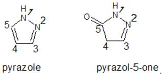
Figure 1. Structures of pyrazole and pyrazol-5-one.
Pyrazolones can be synthesized/ prepared using both the classical and the microwave methods.
The classical method is based on the approach developed by Jensen in 1959 [1]. This is the most common approach, due to its relatively higher yield of products. Again, it was the first method to be known; and more cost effective than the microwave method.
On the other hand, the microwave method involves the irradiation of a mixture of the reactants under microwaves. This method does not provide any significant advantages over the classical/ conventional method in terms of product nature and yield but can reduce the reaction time remarkably avoiding the use of solvent and prolonged heating conditions [2]. Unsubstituted pyrazolones have been synthesized from a hydrazine hydrate and methyl (2E)–3-methoxyacrylate; through the classical method [3], according to the reaction in Figure 2.
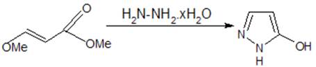
Figure 2. Synthesis of unsustituted pyrazolones.
A solvent-free synthesis of pyrazolones under microwave irradiation has been reported [2]. Here, ethylacetoacetate was made to react with phenyl hydrazine, under microwave irradiation at different power input of 20%, 40% at 2 minutes, 4 minutes in the absence of any solvent. The highest product yield of 82% was obtained with 20% power input at 4 minutes. There was decrease in yield to 55% when the reaction was conducted using increased power input of 40% for 4 minutes. This could be due to the partial decomposition of the product formed. The reaction is represented as shown in Figure 3
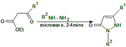
Figure 3. Solvent free synthesis of pyrazolone.
When the classical method was used for the same reaction, carried out in methanol under refluxing condition for ten hours, the product yield was 88% [2].
Pyrazolones belong to the group of organic compound called Heterocyclic compounds. In the recent years they are highly important to chemist due to their pharmacological activities [4, 18-20]. The Pyrazolones in particular have a wide spectrum of biological activity hence they play important role in the design of various bioactive molecules. They possess antimicrobial, antifungal, antimycobacterial, antibacterial, anti-inflammatory, antitumor, gastric secretion stimulatory, antidepressant and antifilarial activities [4,18,20]. The high therapeutic properties of the pyrazolone related drugs have encouraged the medicinal chemists to synthesize a large number of novel chemotherapeutic agents. In line with the medicinal potentials of pyrazolones derivatives, we hereby present synthesis, characterization and anti-prostatic hyperplasia activity of 1-(5-hydroxy-3-methyl-1-phenyl-1h-pyrazol-4-yl) octadecan-1-one.
2. Methodology
2.1. Chemical and Solvents
The chemicals and solvents used in this study were of analytical grade and used as obtained from sigma-Aldrich chemical company without further purification. The chemicals are; β-estradiol-17-valerate and 5α-androstan-17β-ol-3-one (hormone), finasteride (anti-prostatic drug),3-methyl-1-phenylpyrazol-5-one, octadecanoyl chloride, calcium hydroxide. The solvents are ethanol, methanol, toluene, benzene, acetonitrile, tetrahydrofuran, dimethysulfoxide (DMSO), N,N'-dimethylformamide (DMF), acetone, diethylether and petroleum ether.
2.2. Physical Measurements
The melting point was determined using Gallenkamp melting point apparatus. The molar conductance was determined in either DMF using JenwaypeMJ conductivity meter with a cell constant of 1.05. Infrared spectra were collected on Perkin Elmer Paragon 1000 FT-IR spectrophotometer equipped with caesium iodide windo~ (4000-250 cm-1) as KBr pellets. The Proton and Carbon-13 NMR spectra were obtained on a BrukerAvance III HD spectrometer operated at a frequency of 600 MHz in Laboratory F22 of the Department of Chemistry, Rhodes University, South Africa. Dimethylsulfoxide (DMSO) was used as the solvent.
2.3. Synthesis of 1-(5-hydroxy-3-methyl-1-phenyl-1H-pyrazol-4-yl) octadecan-1-one (HMPPO)
1-(5-hydroxy-3-methyl-1-phenyl-1H-pyrazol-4-yl) octadecan-1-one was synthesized according to the procedure outlined in the literature [1], [5-9], with slight modifications. 30g of 3-methyl-1-phenylpyrazol-5-one was dissolved in 140ml dioxane with gentle warming in a 500ml three necked round bottom, quick fit flask equipped with a magnetic stirrer, separating funnel and reflux condenser. 20g of calcium hydroxide was added to form a paste, this was followed by drop wise addition of 20ml octadecanoyl chloride. The mixture was refluxed for about 90 minutes till a light yellow calcium complex was formed. It was allowed to cool. Then 200 ml of 3M HCl were added into the product to decompose the calcium complex whereby a lightyellow coloured crude crystals precipitated and was recrystallized from an ethanol - water (65% - 35%) mixture which was slightly acidified with HCl to decompose any remaining calcium complex and provoke the formation of HMPPO crystals. Equation of reaction is presented in Scheme 1.
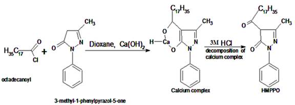
Scheme 1. Equation of reaction for 1-(5-hydroxy-3-methyl-1-phenyl-1H-pyrazol-4-yl) octadecan-1-one (HMPPO).
2.4. Animal Housing
A total of 12 male albino rats having an average weight of about 104.12g each were purchased from an animal breeding unit in the Department of Veterinary Pharmacology, University of Nigeria, Nsuka. The rats were housed in standard steel cages with a plastic base and acclimatized for 7 days under humid tropical condition in the animal house of the College of Natural and Applied Science, Department of Biochemistry, Michael Okpara University of Agriculture, Umudike, Abia State. The rats were exposed to 12hr light/dark cycle and were given free access to clean tap water and commercial rat chow purchased from vital feeds Nigeria Limited.
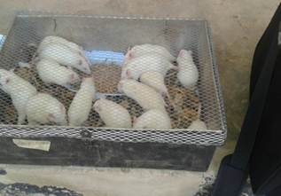
Figure 4. Picture of the experimental rats while in the Biochemistry Department Animal House of Michael Okpara University of Agriculture, Umudike.
2.5. Preparation of the Hormone
This was prepared following literature [10]. 250mg dihydrotestosterone and 25mg estradiol velerate were dissolved in 25ml olive oil and administered intravenously by injection as 9mg/kg body weight estradiol velerate every other day for 28 days. 0.125g of HMPPO was dissolved in 25ml of olive oil to yield a stock of 5mg/ml.
2.6. Experimental Design
After 7 days of acclimatization, the experimental animals were randomly assigned into seven (4) experimental groups of 3 rats each. The groups were labeled as follows.
Group 1 – Standard test control group (STC group); Received no hormone treatment, no HMPPO administration but was fed normal diet for 28 days. This group served as the standard control group.
Group 2 – HMPPO group; Received subcutaneous injection of the hormone DHT and oral administration of the HMPPO everyday for 28 days.
Group 3 – Finasteride group; Received subcutaneous injection of the hormone and oral administration of Finasteride everyday for 28 days
Group 4 – Hormone control group (HC group); Received subcutaneous injection of the hormone every day with no oral administration of the HMPPO or finasteride for 28 days. This group served as hormone control group
2.7. Sample Collection
After 28 days, all the rats were weighed before they were sacrificed by cardiac puncture after dazing with a cervical blow. They were bled exhaustively and the blood was allowed to clot and then centrifuged for 5 minutes at 2000 rpm. The serum was separated and appropriately labeled for analysis. Vital organs for the study were excised and weighed.
2.8. Prostate Specific Antigen (PSA)
This was done following the procedure outlined in literature [11].
2.8.1. Principle
The microwell PSA Enzyme Immunoassay is a solid-phase enzyme immunoassay based on the "sandwich" principle. Two separate antibodies directed against distinct antigenic determinants of PSA molecule are used in this assay. The PSA in the test samples reacts simultaneously with one antibody immobilized on the microwell surface and with another antibody conjugated to horsera dich peroxides enzymes to yield an antibody-antigen-enzyme complex formed on the microwell surface. The unbound conjugate is removed by washing and the colour development reagent (substrate) is added. The intensity of the colour change that occurs reflect the amount of bound anti-PSA enzyme conjugate and is proportional to the concentration of PSA in the sample. The absorbance is read at 450nm.
2.8.2. PSA Procedure
Coated Microwells were set up according to the number of samples to be assayed corresponding to the standard (ST), sample (SA) and control (C). The serum samples (5μl) each and different standards (0.0, 1.0, 2.5, 5.0, 12.5, 25 and 50μ) were added into the Microwell tubes labeled sample (SA) and standard (ST) respectively. A drop of enzyme conjugate was added to each of the samples contained in different wells and mixed thoroughly by rocking for 20 seconds and then sealed with a film sealant. The reaction mixtures were incubated at 37°C for 1hour. The cover sealant was removed and the content of each well was decanted into a sink with disinfectant. Each well was washed with 50μl of the diluted washing buffer (1:9) washing buffer to distilled water). The washing procedure was repeated five times with each washing accompanied by complete drying of the wells by firmly tapping on a pad of tissue paper after discarding the liquid. A drop each of substrate reagent A and substrate reagent B was added to each of the washed wells and gently mixed by rocking for 20 seconds. The mixture was incubated at room temperature for 15 minutes. A drop of stop solution containing 1M hydrochloric acid was added to each of the wells and mixed thoroughly by gently rocking the wells. The well was then loaded in a Stat-Fax-2100 Microwell reader connected to a printer. The solutions were thoroughly mixed before reading their absorbance (A) at 450nm. Earlier, mean values of the absorbance of standards (A) against concentration were plotted to obtain a standard curve from which PSA values were obtained by extrapolation.
2.9. Prostate Acid Phosphate (PACP)
This was done following the procedure outlined in literature [11].
A quantity (1.0 ml) of solution R1was transferred into each of three sets of tubes labeled reagent blank (RB), sample 1 (S1) and sample 2 (S2); while 0.1ml of solution R2 was added to sample 2 (S2) test tube. The reaction media were incubated for exactly 5 minutes at 37°C. the serum sample (0.2 ml) was added to the two sample test tubes labeled sample 1 (S1) and sample 2 (S2) respectively at 30 seconds interval: followed by incubation for 30 minutes at 37°C. after the 30 minutes interval, 10 ml each of diluted sodium hydroxide (NaOH) labeled R3 was added to all the test tubes; followed by the addition of 0.2 ml of the serum sample to reagent blank (RB). The absorbance of reaction medium was read against the blank at 405 nm after being thoroughly mixed.
Note: R1 buffer = citrate buffer, (75 mmol/l, pH5.2), R2 substrate = 1-naphthyl phosphate (10 mmol/l), R3 tartrate = sodium tatrate (135 mmol/l), R4 stabilizer = acetic acid (3 mmol/l).
2.10. Relative Organ Weight Determination
This was calculated as the ratio of the excised organ and the final weight of each rat and the mean taken for each group. Results were expressed as mean ±SD, mean were analyzed for difference using SPSS 17 software.
3. Results
The melting point, colour, yield, conductance and elemental analysis data are listed in Table 1. Solubility data is shown in Table 2. Infrared spectrum, 1HNMR and 13CNMR of HMPPO are presented in Tables 3, 4and 5 respectively. Serum prostate specific antigen (PSA) concentration in rats for the different groups inpresented in Table 6. Table 7 shows Serum prostatic acid phosphatase (PACP) activity concentration in rats for the different groups. Relative prostate weight (RPW) in rats for the different groups is presented in Table 8.
Table 1. Physical and microanalytical data for HMPPO.
| Compound | Molecular Formular | Colour | Yield (%) | Melting point (oC) | Conductance S mol-1cm-1 | Carbon % Found (Calc.) | H % Found (Calc.) | N % Found (Calc.) |
| HMPPO | C28H44N2O2 | Yellowish-brown | 61 | 106 | ….. | 76.21 (76.36) | 10.15 (10.00) | 6.35 (6.36) |
Table 2. Solubility data of HMPPO in various solvents.
| Solvent | HMPPO |
| Chloroform | s |
| Xylene | s |
| Petroleum ether | ss |
| Toluene | vs |
| Methanol | is |
| Cyclohexane | s |
| n-hexane | ss |
| Ethanol | ss |
| DMSO | vs |
| Dioxane | vs |
| Water | is |
Legend: s = soluble, is = insoluble, ss = slightly soluble, vs = very soluble.
Table 3. Selected IR bands of HMPPO.
| Vibration frequency (cm-1) | Assignments |
| 3250.16 | O-H of enol |
| 1743.71 | C=O |
| 3090.07 | Ar C-H |
| 2854.74 | Aliphatic C-H |
| 1618.33 | C=N |
| 1132.25 – 1386.86 | C-H bending |
| 1097.53 | C-H in-plane deformation of the phenyl ring |
Table 4. IH NMR Bands of HMPPO.
| Compound | Phenyl Protons(ppm) | Methyl and methylene protons (ppm) | OH Protons(ppm) |
| HMPPO | 6.84 – 7.42 | 0.48 – 2.15 | 3.65 |
Table 5. 13C NMR Bands of HMPPO.
| Compound | Phenyl Carbons(ppm) | Methyl and methylene carbons (ppm) | C=O(ppm) |
| HMPPO | 120.59 – 158.94 | 13.96 – 33.70 | 174.50 |
Table 6. Serum prostate specific antigen (PSA) concentration in rats for the different groups.
| Groups | No of animals | Mean ± SD (ng/ml) |
| STC | 3 | 0.32±0.04d |
| HMPPO | 3 | 1.18±0.04c |
| FINASTERIDE | 3 | 0.51±0.09c |
| HC | 3 | 2.62±0.34a |
Values are written as mean±sd. values with different superscripts are significantly different (p>0.05) from one another.
Table 7. Serum prostatic acid phosphatase (PACP) activity concentration in rats for the different groups.
| Groups | No of animals | Mean ± SD (ng/ml) |
| STC | 3 | 133.47±1.76 |
| HMPPO | 3 | 339.60±5.07 |
| FINASTERIDE | 3 | 150.72±4.46 |
| HC | 3 | 855.34±8.23 |
Values are written as mean±sd. values with different superscripts are significantly different (p>0.05) from one another.
Table 8. Relative prostate weight (RPW) in rats for the different groups.
| Groups | No of animals | Mean ± SD (g) |
| STC | 3 | 0.29±0.09 |
| HMPPO | 3 | 0.50±0.04 |
| FINASTERIDE | 3 | 0.32±0.02 |
Values are written as mean±sd. values with different superscripts are significantly different (p>0.05) from one another.
4. Discussion
4.1. Microanalytical Measurements
The elemental analysis data in Table 1 shows that HMPPO was prepared. This is as a result of the close percentage values of the carbon, hydrogen, and nitrogen obtained to the calculated value
4.2. Conductivity Measurements
The conductivity measurements of HMPPO in acetone (10-3M) gave no value, indicating its pure covalent nature. The lower the conductivity value, the more covalent a compound is
4.3. Melting Point Determination
The melting point of HMPPO was 106°C. This melting point suggest that HMPPO is non ionic.
4.4. Solubility Data
The solubility data in Table 2 shows that HMPPO is hydrophobic.
4.5. Infrared Spectrum
Infrared spectrum of HMPPO is presented in Table 3. HMPPOshows strong absorption bands around 1132.25 – 1386.86 cm-1 region. This has been assigned to C-H vibrational frequency resulting from bending vibrations in the molecule. The vibrational frequency modes at 1097.53 cm-1have been assigned to C-H in-plane deformation of the phenyl ring. The vibrational frequency 3250.16cm-1has been assigned O-H of enol. The carbonyl functional group appeared at 1743.71 cm-1. Aromatic C-H group functionality was observed at 3090.07 cm-1. Infrared vibration frequencies at 2854.74 and 1618.33 cm-1 were assigned Aliphatic C-H and C=N respectively.
4.6. 1HNMR
The proton NMR spectrum is presented in Table 4. HMPPO showed 1HNMR resonance peak at 3.65 ppm for –OH group. This shows that the enolic form of pyrazolone was prepared. The signals of methyl and methylene protons were observed as multiplets (0.48–2.15 ppm). On the other hand, the signals of the phenyl protons were observed as chemical shifts between 6.84–7.42 ppm in the spectrum of HMPPO.
4.7. 13CNMR
Analyses of the HMPPO13C NMR spectrum (Table 5) indicate that the carbon resonance spectra of C=O was observed at chemical shift value of 174.50 ppm. The Methyl and methylene carbons were assigned 13.96 – 33.70 ppm.
From the elemental analysis, infrared and NMR characterization, the structure in Figure 5 was proposed for HMPPO.
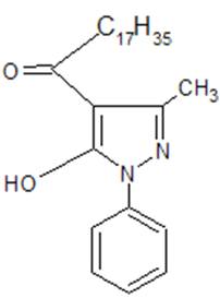
Figure 5. Proposed structure of HMPPO.
4.8. Biological Activity
The result (Table 6) shows a significant increase (P<0.05) in PSA value of ‘HC’ group receiving only hormonal induction with neither HMPPO nor finasteride administration relative to all the other groups. Serum prostatic acid phosphatase activity (Table 7) showed a significant increase (P<0.05) in ‘HC’ Group receiving only hormonal induction with neither HMPPO nor finasteride administration relative to all the other groups. Relative prostate weight was significantly increased (P<0.05) in ‘HC’ group compared to the other groups (Table 8). Relative prostate weight was lowest and significantly different (P>0.05) ‘STC’ and ‘Finasteride’ groups when compared to HMPPO.
Prostate specific antigen is a glycoprotein localized within the prostate gland [12]. It is found in cytoplasmic granules and vesicles, the endoplasmic recticulum, vacuoles and secretory granules, and in lysosomal dense bodies in columnar or cuboidal epithelium of acini but not in acinar cells. It appears to belong to the Kallikrein family of serine proteases [13] with 57% structural similarity. It is 93% protein and 7% carbohydrate of mainly hexose, hexoseamine and sialic acid [14]. PSA is believed to be synthesized in the rough endoplasmic recticulum, stored in the vesicle and vacuole and released in the grandularlumina by exocytosis [11]. Because of its tissue specificity, PSA is used as a marer for BPH and prostate cancer. PSA is a single polypeptide and occurs both in normal and malignant prostatic tissues and in the gland of men with BPH, but not in any other human tissue [11]. This study showed that finasteride reduced significantly (P<0.05) PSA level in the ‘finasteride’ group. Similar observation was also observed for HMPPOwhich reduced PSA significantly (P<0.05) relative to the hormone control ‘HC’ group. The reduction in PSA concentration may have been as a result of decrease in proliferation of prostatic cells in the test groups since PSA level is known to elevate in prostatic lesion due to cellular proliferation.
PSA is a secretory product of prostatic cells and will increase when the prostatic cells increase in number. PSA value can be used to predict enlargement of the prostate because high PSA value correlate increase in prostate mass [15]. This result indicates that HMPPO plays a key role in the chemoprevention of BPH by arresting the proliferation of prostatic cells.
Prostatic Acid Phosphates (PACP) could be used as a marker to detect prostate disorders in human or animals [16]. However, the discovery of PSA has resulted in a shift from PACP to PSA. Elevated PACP level in animals treated with DHT and estradiol and this may be due to hyperplasia of the prostate gland have been reported [17]. Together with PSA, PACP can give useful information about prostatic disease especially BPH prostate cancer. The PACP results of this study are similar to that of PSA. There was a significant increase (P<0.05) in PACP level in the hormone control group (HC group) receiving hormonal induction with neither of HMPPO nor finasteride. It was further observed that PACP level in group 1 was lowest showing that the decrease in PACP in the HMPPO treated groups may have resulted from the HMPPO administration. This finding is consistent with the result of PSA discussed earlier and further indicates that HMPPO may have arrested growth of prostatic cells in the test groups leading to decreased secretion of prostatic acid phosphatase in the prostatic cells.
Benign prostatic hyperplasia results from the enlargement of the prostate gland which causes obstruction of the urethra and the resultant lower urinary tract symptoms. The enlargement of the prostate gland results to increase in the organ weight relative to body weight. The result of this study shows a significant increase (P<0.05) in relative prostate weight of ‘HC’ group compared to the test group HMPPO and Finasteride group. It is important to note that prostate size is the single most important factor in static component of BPH hence any agent that shows positive effect in reduction of prostate size in both normal and diseased prostate may be very therapeutically important in management and prevention of BPH. It is also important to mention that a low urinary output (although not measured but assessed by observation) was observed in the rats in the ‘HC’ group starting from week 3 of the study thus increasing the possibility that the rats in this group may have started experiencing symptomatic BPH in week 3 of this study.
5. Conclusion
The conductivity measurements of the ligand in acetone (10-3M) gave no value, indicating that they are non-ionic compounds. Elemental analysis, infrared spectrum and NMR were used in the characterization of HMPPO. PSA, PACP and prostate size are important markers for benign prostate hyperplasia. The result of this study showed that HMPPO have inhibitory effect on the development of benign prostatic hyperplasia.
References
- Jensen, B.S. (1959). The Synthesis of 1-phenyl-3-methyl-4-acylpyrazolones-5, ActaChemica Scandinavia, 13, 1668–1670.
- Sarbani, P., Jyoti, M. and Nalla, S. D. (2008). High Speed Synthesis of Pyrazolones using Microwave assisted Neat Reaction Technology, J. Braz. Chem. Soc., 19, 1590.
- Eller, G.A. and Holzer, W. (2006). A One-step synthesis of pyrazolone, Molbank, 33: 464.
- Poonam, G.,Jitendra, k., Gupta and halve A. k. (2015). Synthesis and biological significance of pyrazolones: a review. International journal of pharmaceutical science and research. Vol. 6(6): 2291-2310.
- Okafor, E.C., Uzoukwu, A.B., Hitchoock, P.B. and Smith, J.D. (1990). Crystal Structure of bisoxo-bis(1-phenyl-3-methyl-4-acetylpyrazolone-5- O ketoaquouranium(VI), Inorg. Chim. Acta., 172: 97.
- Ogwuegbu, M.O.C. and Maseka, K.K. (1998). Studies on the coordination complexes of Ca (II),Cd (II) and In (IV) with p-nitrobenzoyl-oxo-pyrazole, Bull. Chem. Soc. Ethiop., 12 (1), 27–33.
- Arinze, J.C., Daniel, N.E. and Ogwuegbu, M.O.C. (2012). Synthesis and Characterization of p-nitrobenzoylpyrazolone-5 and its complexes of Mn (II), Fe (III), Rh (III), W (VI) andU (VI), Journal of Emerging Trends in Engineering and Applied Sciences, (JETEAS), 3(1), 61–68.
- Ogwuegbu, M.O.C. (1999). Synthesis and Characterization of Nitroacyl-5-oxo-pyrazole and its Vanadium (V), Iron (III) and Cobalt (II) complexes, Bull. Chem. Soc. Ethiop., 13 (2), 113—120.
- Okafor, E.C. (1984). 1H and 13C NMR Spectral studies of 1-phenyl-3-methyl-4-acylpyrazol-5-ones in chloroform,SpectrochimicaActa.,40(5): 397-401.
- Ahaiwe, U. (2012). The effect of oral administration of leaf extract of VernoniaAmygdalina on benign prostatic hyperplasia in rats. A Thesis of Biochemistry Dept. MOUAU.
- David, A.A. (1993). Prostate – Specific Antigen: Biochemistry Analytical methods and clinical Application. Clin. Chem. 39(2):181–195
- Shina, A.A., Wilson, M.J. and Glesson D.F. (1987). Immunoelectron microscopic localization of prostate specific antigen in human prostate by a protein gold complex.J. Natl.Cancer Inst. Monogr Cancer. 60: 1288–1293.
- Wang, M.C., Valenzuela, L.A., Murphy, G.P. and Chu, T.M. (1981). Purification of a human prostate specific antigen. Invest Urol. 17: 159–163.
- Lilja, H. A. (1980). A killikrein – like serine protease in prostatic fluid cleaves the predominant seminal vesicle protein. J Clin. Invest. 76: 1898–1903.
- De La Rosette, J., Alivizatos, G., Madersbacher, S., Rioja, S.C., Nordling, J., Emberton, M., Gravas, S., Michelm M.C and Oelke, M. (2006). Guidelines on benign prostatic hyperplasia. European Association of Urology. 40: 672–676.
- Joshua, P.E., Obidoa, O. and Nwodo, F.C. (2010). Biochemical responses of rat prostate to coconut (cocosnucifera) milk ingestion/treatment. NJBM. 25(1):1-8.
- Jeyaraj, D.A., Uduyakumar, T.S., Rajalakshmi, M., Pal, P.C. and Sharma, R.s. (2000). Effect of long administration of androgens and estrogen on rhesus monkey prostate: possible induction of benign prostatic hyperplasia. J.Androl. 21:833–841.
- Rani, V.E. and Ravindranath, L.K.(2016). Synthesis and antimicrobial activity of Novel pyrazolo-5-one containing 1,3,4-oxadiazole Sulfonyl Phosphonates, American Journal of organic Chemistry, 6(1): 1–7.
- Prajuli, R. Banerjee, J., Khanal, H. (2015). Synthesis of some pyrazolonederivaties and evaluation of its antibacterial and cytotoxic activity, Orient J.Chem, 31(4):2099-2106.
- Naim, M.J., Alam, O.,Nawaz, F., Alam, A.J. andAlam P. (2016). Current status of pyrazole and its biological activities. J.Pharm Bioallied Sci. 2016, 8(1): 2-17.


