
Chemistry Journal, Vol. 1, No. 4, August 2015 Publish Date: Jun. 17, 2015 Pages: 144-150
The Effect of Acid and Thermal Treatment on a Natural Diatomite
Azimi Pirsaraei Seyed Reza1, Asilian Mahabadi Hasan1, *, Jonidi Jafari Ahmad2, Farahmandkia Zohreh3, Taran Jafar3
1Occupational Health Engineering Department, Faculty of Medical Sciences, Tarbiat Modares University, Jalal Ale Ahmad Highway, Tehran, IR Iran
2Environmental Engineering Department, Faculty of Health, Iran University of Medical Sciences, Shahid Hemmat Highway, Tehran, IR Iran
3Analytical Chemistry & Chemistry Laboratory, Faculty of Health, Zanjan University of Medical Sciences, Parvin Etesaami, Zanjan, IR Iran
Abstract
The characterizations of a natural diatomite such as chemical compositions, specific surface area, total pore volume, pore size distribution and XRD were studied. The diatomite that was treated only with sulfuric acid displayed a larger surface area, expanded total pore volume and increased pore size in comparison with both washed natural diatomite with deionized water (D-H2O) and the diatomite treated with sulfuric acid then calcinated at 550°C (D-H2SO4+550). The specific surface area and total pore volume were increased 46.63% and 71.40%, respectively. The results showed that the diatomite was composed of cristobalite, quartz and feldspar phases. Acid treatment and then calcination at 550°C changed the Si/Al ratio and grew the crystallite size. BJH with DFT analysis showed that the diatomite had very disordered micro/mesoporous pore networks. The pore size distribution of the diatomite was from 1.4-100 nm. Its isotherm was type IV and showed a long hysteresis loop that resembled the H3 type on the basis of the IUPAC classification. Therefore, the diatomite that was treated only with sulfuric acid can be to serve as a catalyst support or an adsorbent.
Keywords
Natural Diatomite, Acid Treatment, X-Ray Diffraction, Crystallite Size Determination, Scanning Electron Microscopy
Received: April 8, 2015
Accepted: April 16, 2015
Published online: June 18, 2015
@ 2015 The Authors. Published by American Institute of Science. This Open Access article is under the CC BY-NC license. http://creativecommons.org/licenses/by-nc/4.0/
1. Introduction
Diatomaceous earth or kieselguhr, also known as Diatomite, is a fine sedimentary rock of biogenetic origin, which mainly consists of amorphous hydrate and silica of diatomite resembles Opal or hydrous silica in composition (SiO2-nH2O). It is usually derived from the skeletons of aquatic plants called diatoms, but it is partly composed of alumina. The bulk of the chemical composition of diatomite (usually 70-90%), is silica and the remains include alumina (0.6-8%), iron oxide (0.2-3.5%), alkali metal oxides, Na2O and MgO (less than 1%), CaO (0.3-3%) and a minor amount of other impurities, such as P2O5, and TiO2. Sometimes, the deposits of the diatoms are only consisting of shell, but it may be contain other sediments such as clay and fine sand. Organic and inorganic materials, carbonates and metals oxides are the typical contaminants. Organic materials and carbonates decompose to CO2 and SO2 gases plus H2O and leave the bulk of diatomite during the thermal operation, such as calcination [1,2]. Diatomite is abundant in many areas of the world and it is easily available in large quantities at an extremely low cost. It has favorable physical characteristics, such as high porosity (25–65%), pore spaces (80-90%), small particles size, low thermal conductivity, low specific gravity range of 1.95 - 2.3, appropriate surface area from 16 to 70 m2 g-1 and high adsorption capacity [3-5].
The important properties of diatomite are related to physical structures and an aggregate of fine particles perforated by a regular pattern of very small holes. The presence of silica can provide the useful characteristics such as a unique structure, chemical stability, and low abrasion. These characteristics are the good causes that the diatomite can become as a filter aid, filler, anti-caking agent, thermal insulator, absorbent, and catalyst carrier. It also can function as a chromatographic support and additive agent for numerous other purposes [5]. Diatomite has been already used for the adsorption of different elements and substances from water and wastewaters, either natural or modified form (chemically or thermally modification). Thermal treatment is expected to cause physical desorption of the adsorbed water from diatomite which belongs to the crystal mesh of the diatomite (forming active hydroxyl groups on it). Generally, thermal treatment has a prominent effect on the type, distribution, content of hydrated species (water, H-bonded silanols, and isolated silanols), and influencing key reactive sites for various surface reactions. Also, thermal treatment can cause to remove the volatile and organic admixtures [6]. Chemical treatment is a common procedure for purifying of diatomite and modifying of its surface properties for various purposes. Typically, the acid activators include sulfuric acid, oxidation with sulfuric acid/H2O2, hydrochloric acid, phosphorous acid and nitric acid. The alkaline activators include sodium hydroxide, sodium carbonate and potassium hydroxide. These are used for purifying diatomite, removing residues and chemically creating finer pores. Acid treatment reduces or eliminates all other oxides of diatomite relative to SiO2 that can become an increase in surface area and adsorption capacity. Alkaline agents such as sodium hydroxide are used to create more open pores. By removing impurities and organic compounds can be expected to gain a larger surface area and total pore volume in diatomite [7-9].
In Iran, diatomite is mainly found in three regional deposits, including the mines of Khorasan (Birjand), Tabriz and the Persian Gulf seashore. The characterizations of the natural diatomite from these deposits were studied by Mahani and Kazemeini (2003). Their study was more focused on the diatomite of the Persian Gulf seashore and Tabriz region but they issued a fewer details about the Birjand diatomite characterizations.
The aim of the present study was to determine the effects of the thermal and acid treatment on characterizations of the Birjand natural diatomite. To serve this purpose, the chemical composition, surface area, pore size and pore volume of the natural diatomite as well as the XRD patterns, the SEM photographs and EDS analysis were determined.
2. Experimental
The natural diatomite contains impurity of the mineral and organic compounds that cause insufficient wettability and low absorption capacity. Also, these impurities can block the pores of the natural diatomite and diminishing usage of it as an adsorbent or catalyst support. Therefore, it is necessary to remove the impurities.
Here, the treatment of the Birjand natural diatomite with sulfuric acid and heat was examined. Before treatment, they were sieved to the size of 8-10 mesh. Fifty grams of it were dispersed in 250 ml of deionized water and then magnetically stirred for 30 minutes. This procedure was repeated several times to remove the impurities. Thus, it was dried in an oven at 110°C for 18 h and weighed again. It was denoted as D-H2O and was kept in plastic bottle until was characterized. A portion of it was used for acid treatment (30g). The acid treatment was performed with 3M H2SO4 solution (Pro-analysis 95-97% sulfuric acid GR for analysis, from Merck) at 100°C for 4 hours in reflux conditions. After cooling at room temperature the slurry was filtered through Whatman No. 42 paper and a Büchner funnel. The diatomite sample was mixed with the acid at a solid (g)/liquid (ml) ratio of 1:20, (30g diatomite/600ml sulfuric acid). The precipitate was repeatedly washed with hot deionized water until the pH levels of the filtered diatomite and the deionized water were the same. Thereafter, the sample was dried at 110 °C for 18 hours, and weighed again. It was named D-H2SO4 and 10g of it was kept in plastic bottle for determining its characterizations. The residues were subjected to the thermal treatment. Thermal treatment was performed under an air atmosphere in a programmable furnace (muffle stove), at heating rate of 10 °C min-1 from room temperature to the calcination temperature. The sample was put in the furnace at 550 °C for 3 hours. It was entitled D-H2SO4+550 and after cooling was weighed again. Having completed the treatments, all of them were subjected to the BET, BJH, XRD, XRF, SEM and EDS analyses. The nitrogen gas adsorption method with Quanta Chrome Nova 2200, version 7.11, high speed gas sorption analyzer was used to measure their specific surface area, along with mean pore size and pore volume distribution. Brunauer-Emmet-Teller (BET) and Barrett-Joyner-Halenda (BJH) methods were also used for determining the specific surface area and the total pore distribution, respectively. For ascertaining the crystal structure of them, X-ray diffraction analysis (XRD) was performed using Philips X'Pert equipment with a cobalt tube anode diffractometer (λ= 0.178897 nm) operated at 40 kV and 40 mA in a continuous scan mode by scanning range of 3-70° (2θ) at 0.8s per step. The X-ray fluorescence technique (XRF) was done for elemental analysis. The morphology of the diatomite was considered using a scanning electron microscopy (SEM, VEGA\\TESCAN) equipped with an energy dispersive X-ray spectroscopy (EDS).
3. Result and Discussion
3.1. XRD and XRF Analysis
The XRD patterns of the diatomite are given in fig. 1. The Birjand natural diatomite was essentially amorphous, however contained quartz, cristobalite, feldspar, as well as small amounts of illite, magnesiohornblende and montmorillonite (clay).
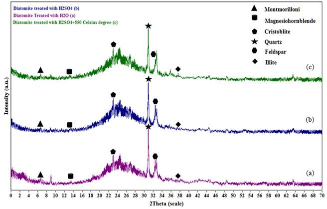
Fig. 1. XRD patterns of the diatomite: (a) D-H2O, (b) D-H2SO4, (c) D-H2SO4+550.
The X-ray diffraction spectra displayed that silica was the major component. The cristobalite, quartz and feldspar noteworthy were revealed in the range of 25< 2θ <33 in the X-ray diffraction spectra, respectively.
(table 1) shows elemental analysis of the diatomite before and after treatments, obtained by XRF technique. According to the results of analysis, the Birjand natural diatomite had less impurities and higher silica content (>82 wt %). The main chemical compositions were the oxides of Si and Al. The other important components were the metal oxides of Fe, K, Ca, and Mg.
Table 1. The chemical characterization of the diatomite.
| Chemical content (% weight) | Natural Diatomite | D-H2O | D-H2SO4 | D-H2SO4+550 |
| SiO2 | 82.16 | 82.27 | 89.98 | 92.17 |
| Al2O3 | 4.89 | 4.97 | 2.58 | 2.82 |
| Fe2O3 | 1.46 | 1.81 | 0.39 | 0.32 |
| CaO | 1.23 | 1.47 | 0.72 | 0.82 |
| MgO | 0.87 | 1.29 | 0.24 | 0.21 |
| MnO2 | N.D.ä | N.D.ä | N.D.ä | N.D.ä |
| K2O | 0.54 | 0.90 | 0.53 | 0.64 |
| Na2O | 0.43 | 0.54 | 0.42 | 0.45 |
| TiO2 | 0.19 | 0.23 | 0.09 | 0.15 |
| P2O5 | 0.12 | 0.17 | 0.02 | 0.00 |
| L.O.I. | 8.07 | 6.27 | 4.90 | 2.26 |
äN.D. = Not detected
The results indicated that the acid treatment procedure has increased silica content and leached out organic and inorganic impurities. However, the thermal treatment method at 550 °C has increased silica content, which may be attributed to crystallite growth after heating and slow cooling along with more removal of the impurities. Also, the loss on ignition (L.O.I.) was the lowest percentage in D-H2SO4+550, than D-H2O and D-H2SO4. It showed that the organic compounds were removed by heating at a temperature of 550 °C.
The crystallite size broadening (β) of a peak can usually be related to the crystallite size (L) via the Scherrer [10,11]. This is because of the crystallite size and is not synonymous with particle size, while X-ray diffraction is sensitive to the crystallite size inside the particles. From the well-known Scherrer formula the average crystallite size, L, is:
![]()
Where λ is the X-ray wavelength in nanometer (nm), β is the peak width of the diffraction peak profile at half maximum height resulting from small crystallite size in radians and K is a constant related to crystallite shape, normally taken as 0.9. The value of β in 2θ axis of diffraction profile must be in radians. Determination of crystallite size by XRD is ideal when crystallites are too small for optical methods, and XRD provides a bulk material sampling [12,13]. In our study, according to the respective X-ray diffraction data, the crystallite size of the three crystalline phases (quartz, cristobalite and feldspar) was decreased through acid treatment. However, the crystallite size was increased due to a nucleating and crystalline growth at 550°C calcination temperature (see table 2).
Table 2. X-ray diffraction data for the diatomite.
| D-H2O | D-H2SO4 | D-H2SO4+550 | |||||||
| d (Å) | FWHM (degree) | Crystallite size (nm) | d (Å) | FWHM (degree) | Crystallite size (nm) | d (Å) | FWHM (degree) | Crystallite size (nm) | |
| Cristobalite | 4.06 | 1.5605 | 6.57 | 4.08 | 2.2086 | 4.53 | 4.06 | 0.6311 | 18.51 |
| Quartz | 3.35 | 0.1328 | 771.40 | 3.35 | 0.1751 | 174.98 | 3.35 | 0.1605 | 238.67 |
| Feldspar | 3.18 | 0.1480 | 348.13 | 3.18 | 0.1664 | 208.91 | 3.19 | 0.1384 | 533.61 |
3.2. SEM with EDS Analysis
The SEM micrograph structure and EDS analysis of the diatomite are given in fig. 2. The pores are observable on the surface of the diatomite (see fig. 2a). After the diatomite was treated with H2SO4, the pores were preserved and slightly opened, but some changes were perceived in the diatomite structure after calcination at 550 °C for 3 h (see fig. 2b and c). The SEM images of D-H2O, D-H2SO4 and D-H2SO4+550 showed a difference between their surface textures and pores. According to the EDS elemental analysis, silica was the main element (see fig. 2d, e and f) and the EDS results indicated that the Si/Al ratio for D-H2O, D-H2SO4 and D-H2SO4+550 were 21.11, 20.05 and 25.01, respectively.
3.3. BET Surface Area and BJH Analysis
The results obtained from the specific surface area (BET) and pore volume distribution (BJH and DFT) of the diatomite are presented in (table 3 and 4). The specific surface area (BET), total pore volume and average pore radius of D-H2O were 29.14 m2g-1, 0.0432 cm3 g-1 and 29.65Å, respectively. When the diatomite was only treated with sulfuric acid, an increase in the BET surface area (~ 29 m2g-1 for D-H2O to ~ 43 m2g-1 for D-H2SO4), the total pore volume and average pore radius was appeared.
After being treated with H2SO4, the surface area of the diatomite increased up to 46.63%, and its porosity was improved significantly. When this diatomite was subjected to 550 °C for 3 h (D-H2SO4+550), the specific surface area decreased dramatically. This treatment was resulted in a decrease in the total pore volume of the diatomite, while the average pore volume was increased (see table 3 and 4). Therefore, a combination of chemical and thermal treatment on the diatomite was led to decrease in specific surface area and total pore volume. These structural changes were also observed by Aivalioti et al [2,9]. According to IUPAC, the shape of the adsorption isotherm can be classified into one of six groups [14-17]. Fig. 3(a) shows that the adsorption volume at low relative pressures (p/p0< 0.40) was positive due to the presence of micropores. Increasing the relative pressure had caused the capillary condensation which was illustrative of type IV behavior.
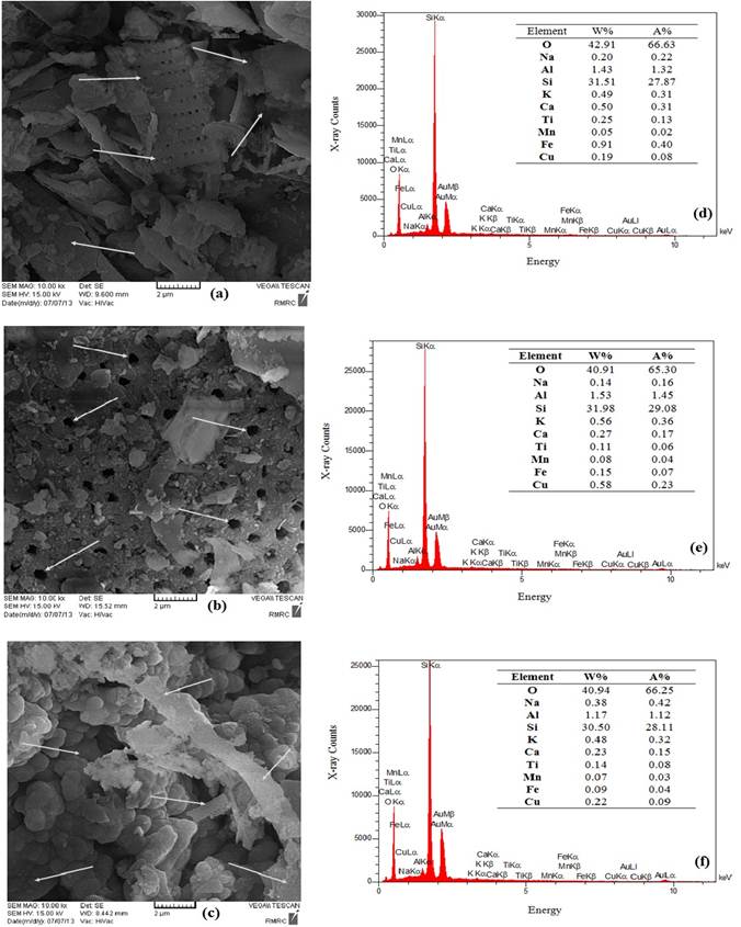
Fig. 2. SEM images (magnification 10000×) and EDS analysis: (a) diatomite washed with deionized water and dried at 110 °c for 18 h (D-H2O), (b) diatomite treated with H2SO4 (D-H2SO4), (c) diatomite treated with H2SO4 and then calcinated at 550 °c (D-H2SO4+550), (d) EDS of D-H2O, (e) EDS of D-H2SO4 and (f) EDS of D-H2SO4+550, respectively.
Table 3. The physical characterization of the diatomite.
| Sample | Specific Surface area (BET) (m2 g-1) | Total Pore volume (cm3 g-1) | Average pore radius (Å) |
| D-H2O | 29.14 | 0.04319 | 29.65 |
| D-H2SO4 | 43.31 | 0.07403 | 34.19 |
| D-H2SO4+550 | 16.81 | 0.03492 | 41.55 |
Table 4. The microspores and mesopores characterization of the diatomite.
| Sample | Micropore volume (cm3 g-1) | Micropore area (m2 g-1) | Mesopore volume (cm3 g-1) | Mesopore area (m2 g-1) | Micropore content (%) | Mesopore content (%) | Pore width (Å) | |
| D-H2O | 0.0078 | 16.63 | 0.0354 | 12.51 | 18.06 | 81.94 | 4.17 | |
| D-H2SO4 | 0.0101 | 14.85 | 0.0639 | 28.46 | 13.64 | 86.36 | 17.64 | |
| D-H2SO4+550 | 0.0039 | 5.58 | 0.0311 | 11.23 | 11.17 | 88.83 | 16.87 | |
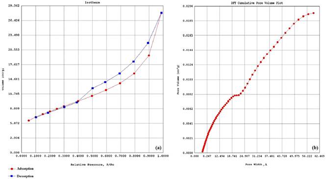
Fig. 3. (a) A typical of nitrogen adsorption (circle symbols) and desorption (rectangular symbols) isotherm for the diatomite and (b) pore size distribution curve (Density Functional Theory=DFT) for micropores/macropores measurement.
Therefore, the isotherm type of the diatomite was the type IV. In our study, the adsorption–desorption isotherm of the diatomite (nitrogen gas adsorption method by a Nova 2200 Quanta Chrome analyzer) showed the long hysteresis loop that was look like to the H3 type in the IUPAC classification (see Fig. 3(a) a typical long hysteresis loop). In fact, the H3 type indicated the presence of very disordered micro/mesoporous pore networks were caused by a combination of various phenomena, including cavitation and pore blocking mainly due to pore cavity size from adsorption branch. On the basis of the adsorption branch of the isotherm, the pore size distribution of the diatomite was from 14 Å to 1000Å (1.4–100 nm).
Fig. 3 (b) shows a DFT isotherm that is used for the merged micro-mesoporous analysis. At higher pressures, the slope showed an increase in uptake of adsorbate because the pores were clogged. The inflection point typically occurred near completion of the first monolayer. In this diatomite type, the mesopores were dominant.
4. Conclusion
The conclusion of data related to the surface area and the pore size from the treated diatomite only with acid indicated that the pores structures remained unchanged while the blocked pores were opened.
Finally, the following major conclusions were drawn:
• Since the isotherm type of this diatomite is type ΙV and the hysteresis loop is similar to the H4 type according to the IUPAC classification, the diatomite is likely proper to take for adsorption of chemical compounds.
• Treatment the diatomite with 3M H2SO4 (in this study so-called D-H2SO4) creates a fairly high BET surface area and an increase in total pore volume. Therefore, the diatomite can be introduced as a proper solid matter for impregnating with chemical agents.
• Diatomite is an inexpensive source of amorphous hydrated silica with the cristobalite, quartz and feldspar crystalline phase. Its major chemical components are SiO2, Al2O3 and Fe2O3. The presence of these compounds can improve the diatomite mechanical strength.
• Diatomite is an inert support that has positive features for catalytic active components. Also its natural pores and mesh-like openings can reduce the need for porosity producing agents during the catalyst preparation.
Acknowledgement
The financial support of this research was conducted by Research Deputy of Tarbiat Modares University. The authors thank Professor M. Khamechian, Mrs. Z. Fardindoost and Mr. M. Yousefi in Geology Department Laboratory, Tarbiat Modares University for help to the XRD analysis. The authors are appreciative of Mrs. Ziba Hasanvand and her colleagues the Iran Polymer & Petrochemical Institute, for BET & BJH analysis. We are grateful Mr. Dehghani from the Razi Metallurgical Research Center for SEM photography. Also the authors thank Mr. S. Sabouri Raad for obtaining the South Khorasan diatomite.
Abbreviations
| Full phrase | Abbreviation |
| Barrett-Joyner-Halenda | BJH |
| Brunauer-Emmet-Teller (surface area) | BET |
| Density Functional Theory | DFT |
| Washed diatomite with deionized water | D-H2O |
| Modified diatomite with sulfuric acid | D-H2SO4 |
| Modified diatomite with sulfuric acid and then calcined at 550 °C for 3 h | D-H2SO4+550 |
| Energy dispersive X-ray spectroscopy | EDS |
| International Union of Pure and Applied Chemistry | IUPAC |
| Scanning electron microscopy | SEM |
| X-ray diffraction | XRD |
| X-ray fluorescence | XRF |
References
- M.B. Morsy Heg. Diatomite: Its Characterization, Modifications and Applications.Asian J. Mater. Sci. 2010; 2: 121-136.
- M. Aivalioti, I. Vamvasakis, E. Gidarakos.BTEX and MTBE adsorption onto raw and thermally modified diatomite. J. Hazard. Mater. 2010; 178: 136-143.
- P. Vassileva, G. Gentscheva, E. Ivanova, P. Tzvetkova, D. Voykova, M. Apostolova.Characterization of natural diatomites from Bulgaria. Compt. Rend. Acad. Bulg. Sci. 2011; 64: 823-830.
- H. Mahani, M. Kazemeini. Treatment of iatomaceous earth to obtain its catalyst support. Sci. Iranica. 2003; 10: 350-356.
- S. Dehestani Athar, H. Asilian.Catalytic oxidation of carbon monoxide using copper-zinc mixed oxide nanoparticles supported on diatomite. J. Health Scope. 2012; 1:52-56.
- A. Chaisena, K. Rangsriwatananon. Effects of thermal and acid treatments on some physic-chemical properties of Lampang diatomite. Suranaree. J. Sci. Technol. 2004; 11: 289-299.
- P.S. Vassileva, M.S. Apostolova, A.K. Detcheva, E.H. Ivanova.Bulgarian natural diatomites: modification and characterization. Chem. Pap. 2013; 67: 342–349.
- G. Zhang, D. Cai, M. Wang, C. Zhang, J. Zhang, Z. Wu. Microstructural modification of diatomite by acid treatment, high-speed shear, and ultrasound. Microporous Mesoporous Mater. 2013; 65: 106-112.
- M. Aivalioti, P. Papoulias, A. Kousaiti, E. Gidarakos.Adsorption of BTEX, MTBE and TAME on natural and modified diatomite. J. Hazard. Mater. 2012; 208:117-127.
- R. Jenkins, R.L. Snyder. Introduction_to_X-ray powder diffractometry. J D Winefordner series editor. John Willey & Sons, New York, 1996, pp. 47-94.
- E. Lifshin. X-ray characterization of materials. Weinheim: WILEY-VCH Verlag GmbH Germany, D-69469, 1999, pp. 1-105.
- A. Monshi, M.A. Foroughi, M.A. Monshi.Modified Scherrer equation to estimate more accurately nano-crystallite size using XRD. World. J. Nanoscience. Eng. 2012; 2: 154-160.
- V. Victor Drits, J. Srodon, D.D. Eberl. XRD measurement of mean crystallite thickness of Illite and Illite/Smectite: Reappraisal of the Kubler index and the Scherrer equation. Clays Clay miner. 1997; 45: 461-475.
- K.S.Sing, D.H. Everett, R.A.W. Haul, L. Moscou.R.A. Pierotti, J. Rouquerol, T. Siemieniewska.Reporting physisorption data for gas/solid systems with special reference to the determination ofsurface area and porosity.Pure Appl. Chem. 1985; 57: 603-619.
- P.B. Balbuenat, K.E. Gubbins. Theoretical interpretation of adsorption behavior of simple fluids in slit pores.Langmuir. 1993; 9: 1801-1814.
- S. Naumov.Hysteresis phenomena in Mesoporous materials [dissertation].Faculty of physics and Geosciences university of Leipzig, 2009.
- M. Thommes. Physical adsorption characterization of nanoporous materials. Chem. Ing. Tech.2010; 82: 1059-1073.


