
Public Health and Preventive Medicine, Vol. 1, No. 2, June 2015 Publish Date: May 28, 2015 Pages: 73-77
Detection of Naegleria Isolates from the Egyptian Aquatic Environment
Wafaa M. Hikal1, 2, *, Ahmad Z. Al-Herrawy2, Mahmoud M. Bahgat3, Abd-Elhafez H. Mohammed4, Ameen A. Ashour4
1Department of Biology, Faculty of Science, University of Tabuk, Tabuk, Saudi Arabia
2Parasitology Lab, Water Pollution Research Department, National Research Centre, Dokki, Giza, Egypt
3Therapeutic Chemistry Department, National Research Centre, Dokki, Giza, Egypt
4Zoology Department, Faculty of Science, Ain Shams University, Cairo, Egypt
Abstract
Free-living amoebae of the genus Naegleria have been recognized as etiologic agents of amoebic encephalitis, keratitis, otitis, lung lesions and other skin infections mainly in immuno-compromised individuals. Naegleria fowleri is the causative agent of primary amoebic meningo-encephalitis (PAM), a rapidly fatal disease of the central nervous system. The disease is generally acquired while swimming and diving in freshwater. In the present study samples from swimming pools water in Egypt were examined for Naegleria using a polymerase chain reaction (PCR) method. Members of genus Naegleria were detected in 27.5% of the examined swimming pool water samples. Based on the morphological attributes of trophozoites and cysts, flagellation test, all the isolates were classified to the genus Naegleria. Molecular identification of the amoebae isolated from water samples confirmed their affinity to Naegleria genus. The isolated species of Naegleria could provoke variable degrees of infections to the swimmers. Thus there is a need for further investigation to establish Naegleria genotype.
Keywords
Free-Living Amoebae, Naegleria, Flagellation Test, PCR, Swimming Pools
Received: April 8, 2015
Accepted: April 25, 2015
Published online: May 27, 2015
@ 2015 The Authors. Published by American Institute of Science. This Open Access article is under the CC BY-NC license. http://creativecommons.org/licenses/by-nc/4.0/
Contents
1. Introduction 2. Materials and Methods 2.1. Samples and Sampling Sites 2.2. Isolation and Morphologic Identification of Naegleria Spp from Water Samples 2.3. Flagellation Test 2.4. Molecular Characterization of Isolated Freshwater Amoebae Using Polymerase Chain Reaction (PCR) 3. Results 3.1. Prevalence of Naegleria in the Examined Swimming Pools 3.2. Morphological Characterization of Genus Naegleria 3.3. PCR Product of Genus Naegleria 4. Discussion 4.1. Prevalence of Heat Tolerant Free-Living Amoebae in Different Types of Water 4.2. Morphological Characterization of Genus Naegleria 4.3. Molecular Characterization of Genus Naegleria 5. Conclusion
1. Introduction
Naegleria is a free-living amoeba that is ubiquitously distributed in the environment worldwide in fresh water as well as in marine water cooling towers (Barbaree et al., 1986),. Moreover, they have been recovered from various domestic water systems such as drinking tap water (Michel et al., 1998).Only Naegleria fowleri has been shown to cause human disease, that result in primary amoebic meningo-encephalitis (PAM), a rapid fatal infection of the central nervous system (CNS) that occurs generally in previously healthy children and young adults with a history of exposure to contaminated recreational, domestic, or environmental water sources (Ithoi et al 2011).
More than 200 cases of PAM, predominantly in children and young adults, were reported worldwide as of July 2002 (Visvesvara, 2003).
Naegleria fowleri (N. fowleri) feed on red blood cells, white blood cells, and brain tissue. PAM is almost fatal, usually killing its victims within 3–7 days after the onset of symptoms (CDC, 2010; Cabanes et al, 2001; John, 1982).Pathogenic FLA are not dependent upon a host for transmission and spread, nor does host-to-host transmission of these amoebic diseases occur. They feed by phagocytosis, mainly on bacteria, fungi and algae (Bass and Bischoff, 2001).They have the ability to multiply and grow well in tropical climate and in water body with high temperatures of 40-45ºC (De Jonckheere, 2006).Molecular methods such as PCR offer an attractive alternative to microscopy and culture, since they can be performed by personnel without a high level of expertise in recognizing diagnostic morphological features of amoebae. Furthermore, molecular methods are very sensitive and may allow the detection of fewer microorganisms per volume of sample analyzed than morphological methods would. The aim of this study was to determine the presence of Naegleria spp. in different aquatic environment of Egypt using morphological and molecular characterization methods which can be a risk factor for people especially contact lens wearers and immuno-compromised patients.
2. Materials and Methods
2.1. Samples and Sampling Sites
Water samples (1 liter volume each) were collected monthly from ten different swimming pools in Cairo, Egypt for one year period. Samples were collected in clean, dry autoclavable polypropylene containers and sent to the laboratory of parasitology, water pollution Research Department, National Research Centre, in icebox and processed at the same day of collection.
2.2. Isolation and Morphologic Identification of Naegleria Spp from Water Samples
Collected swimming pool-water samples were separately concentrated by using the membrane filtration technique. One liter of each water sample was filtered through a ni-trocellulose membrane filters (0.45μm pore size and 47mm in diameter) (Whatman, WCN type, Cat No. 7141-104) (Gradus et al., 1989; Hikal, 2010). After filtration the membranes were separately inverted face to face on the surface of a non-nutrient (NN) agar plates previously seeded with 100 μl Escherichia coli suspension. All the inoculated plates were incubated at 40°C for one week with daily microscopic examination for the presence of any amoebic growth (Hikal, 2015). Identification of the obtained Naegleria spp. were achieved according to the morphological characteristics of both trophic and cyst stages (Pussard and Pons, 1977, Hikal, 2010, Al Herrawy et al., 2013).
2.3. Flagellation Test
The obtained amoebic trophozoites were gently scraped from the surface of agar plates with a bacteriological loop and suspended in a test tube containing 5 ml distilled water and incubated at 37°C for 30 minutes. Every 10 minutes one drop from the content of the tube was suspended in the concavity of a clean glass hanging drop slide and examined under the microscope for the formation of temporary flagella (Behets et al., 2003).
2.4. Molecular Characterization of Isolated Freshwater Amoebae Using Polymerase Chain Reaction (PCR)
2.4.1. DNA Extraction
The amoebae pellet was resuspended in lysis buffer containing 2% CTAB as described by Winnepenninckx et al.(1993) and modified by Abdel-Hamid et al. (1999), overlaid with 500 ml of phenol-chloroform-isoamylalcohol (PCI), and shaken gently for 5 hr. The suspension was centrifuged at 3000 xg for 10 min, and the upper, aqueous phase was transferred to a new tube. PCI extraction was repeated two times for 10 min each time. DNA was precipitated at -80°C overnight, pelleted at 12000 xg for 30 min at 4°C, washed in 70% ethanol, air dried, and re-suspended in 30 ml of sterile double-distilled water (Walochnik et al., 2000).
2.4.2. Polymerase Chain REACTION (PCR)
For molecular identification, the genus specific primers were used. Forward primer sequence (5 TTTGAATTCGCTCC-AATAGCGTATATTAA-3) and Reverse primer (5- TTTCTT-TTCCTCCCCTTATTA-3(ٓ (Pelandakis et al. 2000). All amplification reactions of PCR were performed in a 50 μl. PCR consisted of 1 min denaturation at 94°C, 1min annealing at 47°C and 1 min elongation at 72 °C for 35 cycles.After that, 10min of extension time at 72 °C was done. Finally, the PCR products were cheeked by electrophoresis in a 1.5 % agarose gel (Helling et al., 1974).
3. Results
3.1. Prevalence of Naegleria in the Examined Swimming Pools
Naegleria species were detected in 33 (27.5%) water samples collected from 10 swimming pools in Cairo (Table 1).
Table 1. Prevalence of Naegleria spp. in swimming pool samples
| Swimming pools | Examined samples (n) | Naegleria spp. | |
| Number | % | ||
| 1 | 12 | 2 | 16.7 |
| 2 | 12 | - | - |
| 3 | 12 | 4 | 33.3 |
| 4 | 12 | - | - |
| 5 | 12 | 7 | 58.3 |
| 6 | 12 | 8 | 66.7 |
| 7 | 12 | 1 | 8.3 |
| 8 | 12 | 6 | 50.0 |
| 9 | 12 | 3 | 25.0 |
| 10 | 12 | 2 | 16.7 |
| Total | 120 | 33 | 27.5 |
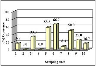
Figure 1. Occurrence of Naegleria spp. in swimming-pool samples
Swimming pool number 6 showed the highest incidence of heat-tolerant Naegleria species (66.7%).The heat-tolerant Naegleriaspecies were not recorded in water samples collected from swimming pool number 4 and 2 (Table 1, Fig. 1).
3.2. Morphological Characterization of Genus Naegleria
Genus Naegleria represented the amoebo-flagellates whose members could transform from amoebae to flagellate forms. The life cycles included amoeboid and cystic stages and for most species a transient flagellate stage. Differentiation of Naegleria from other amoebae was based on their characteristic eruptive movement of the amoebic form, and their ability to transform to flagellates (Figure 2 [{A, B}).
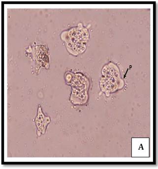
Trophozoite form
Cyst form
Figure 2.Trophic and cystic form of Naegleria
3.3. PCR Product of Genus Naegleria
87.9% of microscopically Naegleria +ve swimming pool samples were also +ve by PCR technique. Microscopically Naegleria +ve swimming pool samples collected from site 1 (n=2), site 5 (n=7), site 8 (n=6) and site 9 (n=3) were all +ve by PCR. It was also observed that 87.5%, 75.0 and 50.0% of microscopically Naegleria +ve swimming pool samples collected from sites 6, 3 and 10, respectively proved to be +ve by PCR.Electrophoresis of amplification products from ITS primers of different Naegleria isolates were subjected to electrophoresis on 1.5% agarose gel parallel containing ethidium bromide to 100 bp DNA ladder where 409 bp specific amplification products were visualized in all environmental samples tested that were not evidenced in the negative control (Figure 3).
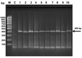
Figure 3. Electrophoresis of amplification products from DNA of different isolates of Naegleria (1, 2, 3, 4, 5, 6, 7, 8, 9 and 10): N. spp., were subjected to electrophoresis on 1.5% agarose gel parallel containing ethidium bromide to M: 100 bp DNA ladder, where 409 bp specific amplification products were visualized in all samples. C: negative control bacteria
4. Discussion
The present study deals with the natural distribution of members of the genus Naegleriain the examined swimming pool water of Cairo, Egypt. To the best of our knowledge, few studies were conducted reporting the detection and existence of Naegleriain Egypt (Hikal, 2010;Al-Herrawy, 2014).
4.1. Prevalence of Heat Tolerant Free-Living Amoebae in Different Types of Water
Free-living amoebae were isolated at 37°C from 73.3% of the examined swimming pool samples. In Egypt, a lower incidence of free-living amoebae (32%) in swimming pools (Hamadto et al., 1993). Other workers in Poland detected free-living amoebae in 59.7% of the examined swimming pool samples (Gronik and Kuzna-Grygiel, 2004). Free-living amoebae grown at 40°C were isolated from 60% of the swimming pool samples. In Poland, Gronik and Kuzna-Grygiel, (2004) recorded a lower incidence of free-living amoebae (37.2%) isolated at 42°C from swimming pools.
4.2. Morphological Characterization of Genus Naegleria
In the present study, it was found that a trophozoite of Naegleria was long slender or oval, measuring 12-35µm in length and 10-30µm in width. Naegleria trophozoites were also characterized by a single vesicular nucleus having a large prominent centrally located nucleolus and a single broad hemispherical hyaline eruptive lobose pseudopodium. These findings are in agreement with those of most other workers (Page, 1974; Marciano-Cabral, 1988; Al-Herrawy, 1992; Ashmawy et al., 1993; Al-Herrawy and Al-Rashied, 1995; Schuster and Visvesvara, 2004; Shin and IM, 2004). In the present study, the flagellate stage of Naegleria amoebae usually had one pair of equal flagella arising from the pointed anterior end. Previous authors reported that the main process was the formation of one pair of flagella per cell (Page, 1967; Visvesvara, 1980; John, 1982; Ashmawy et al., 1993).
In the present study, the cyst form of Naegleria had a hardly detectable double wall with 4-5 shallow pores. These findings are in agreement with these of Schuster and Visvesvara (2004). Naegleria species were too similar morphologically to be distinguished from each other at the level of the ordinary light microscope. This conclusion was also reached by De-Jonckeree (1977) and Ashmawy et al. (1993).
4.3. Molecular Characterization of Genus Naegleria
In the present study the morphologically identified free-living amoebae belonging to the genus Naegleria were confirmed by PCR using genus-specific primers.
The incidence of Naegleria spp. were molecularly detected in 87.9% out of 33 morphologically Naegleria +ve samples (i.e. 27.5% of the total examined) collected from swimming pools. Based on the PCR amplification with a genus-specific primer pair in Taiwan Hsu et al. (2009) detected Naegleria spp. in 5.9% from swimming pool samples.
5. Conclusion
Swimming pools water may be the source of Naegleriainvasion. The use of molecular methods to identify free-living amoebae of genus Naegleria could provide a more rapid means to diagnose infections caused by those amoebae.There is a need for further investigation to establish Naegleria genotype.
References
- Abdel-Hamid, A. Z., Molfetta, J. B., Fernandez, V. and V. Rodrigues (1999). Genetic variation between susceptible and non-susceptible snails to Schistosoma infection using random amplified polymorphic DNA analysis (RAPDs). Rev. Inst. Med. Trop. Sp., 41: 291-295.
- Al-Herrawy, A., Bahgat, M., Mohammed, A., Ashour, A. andW. M. Hikal (2013). Morpho-Physiological and Biochemical Criteria of Acanthamoeba spp. Isolated from the Egyptian Aquatic Environment. Iranian J. Parasitol., 8 (2): 302-312.
- Al-Herrawy, A., Bahgat, M., Mohammed, A., Ashour, A. and W. M. Hikal (2013). Morpho-Physiological and Biochemical Criteria of Acanthamoeba spp. Isolated from the Egyptian Aquatic Environment. Iranian J. Parasitol., 9(2): 194-201.
- Al-Herrawy, A. Z. (1992). In vitro cultivation of agents of amoebic meningo-encephalitis isolated from water and sewage. Ph. D. thesis, Fac. Vet. Med., Alexandria Univ., Egypt.
- Al-Herrawy, A. Z. and K. A. S. Al-Rasheid (1995). Isolation of thermo-tolerant Naegleria from a freshwater course in Riyadh (Saudi Arbia). J. Vet. Sci., 1: 79-83.
- Ashmawy, K., Hilali, M., Abu El-Wafa, S. A., Samaha, H., Draz, A. A. and A. Salem (1993). In vitro identification of Naegleria and Acanthamoeba isolated from water and sewage. Dept. Pathol. Parasitol., 30: 87-98.
- Barbaree, J. M., Fields, B. S., Feeley, J. C., Gorman, G. W. and W. T. Martin (1986). Isolation of protozoa from water associated with a legionellosis outbreak and demonstration of intracellular multiplication of Legionella pneumophila. Appl. Environ. Microbiol., 51: 422-424.
- Bass, P. and P. J. Bischoff (2001). Seasonal variability in abundance and diversity of soil gymnamoebae a short transect in southeastern USA. J. Eukaryot. Microbiol., 48: 475-479.
- Behets, J., Seghi, F., Declerck, P., Verelst, L., Duvivier, L., Van Damme, A. and F. Ollevier (2003). Detection of Naegleria spp. and Naegleria fowleri: a comparison of flagellation tests, ELISA and PCR. Wat. Sci. Technol., 47: 117-122.
- Cabanes, P., Wallet, F., Pringuez, E. and P. Pernin (2001). Assessing the Risk of Primary Amoebic Meningoencephalitis From Swimming in the Presence of Environmental Naegleria fowleri. Applied & Envir. Microbiol., 67:7:2927.
- CDC (Centers for Disease Control and Prevention), 2010.www.dpd.cdc.gov/dpdx/HTML/FreeLivingAmebic.htm(accessed Oct. 31, 2011).
- De Jonckheere, J. F. (2006). Molecular identification of free-living amoebae of the Vahlkampfiidae and Acanthanoebidae isolated in Arizona (USA). Eur. J. Protistol., 43: 9–15
- De Jonckheere, J. F. (1977). Use of an axenic medium for differentiation between pathogenic and non-pathogenic Naegleria fowleri isolates. Appl. Environ. Microbiol., 33: 751-757.
- Gradus, M. S., Koenig, S. B., Hyndiuk, R. A. and J. De Carlo (1989). Filter-culture technique using amoebae saline transport medium for the noninvasive diagnosis of Acanthamoeba keratitis. Am. J. Clin. Pathol., 92: 682-685.
- Gronik, K. and W. Kuzna-Grygiel (2004). Presence of virulent strains of amphizoic amoebae in swimming pools of the city of Szczecin. Ann. Agric. Environ. Med., 11: 233-236.
- Hamadto, H. H., Aufy, S. M., El-Hayawan, I. A., Saleh, M. H. and I. M. Nagaty(1993). Study of free-living amoebae in Egypt. J. Egypt. Soci. Parasitol., 23: 631-637.
- Helling, R. B., Goodman, H. M. and H. W. Boyer (1974). Analysis of R. EcoRI fragments of DNA from lambdoid bacteriophages and other viruses by agarose-gel electrophoresis. J. Virol., 14: 1235-38.
- Hikal, W. M. (2010). Biochemical and molecular characterization of pathogenic free-living amoeba in the aquatic environment. Ph. D. thesis, fac. Sci., Ain Shams Univ., Egypt.
- Hikal, W. M. (2015). Detection of Acanthamoeba Species from Water Tanks in Saudi Arabia. Asian Academic ResearchJ. Multidisciplnary, (1): 42-49.
- Hsu, B., Lin, C. and F. Shine (2009). Survey of pathogenic amoebae and Legionella spp. in Mud Spring recreation area. EGU General Assembly 2009, Vienna, Austria.
- Ithoi, I., Ahmad, A. F., Nissapatorn, V., Lau, Y. L.; Mahmud, R. and J. W. Mak (2011). Detection of Naegleria species in environmental samples from Peninsular Malaysia. PLoS One, 6:e24327.
- John, D. T. (1982). Primary amoebic meningo-encephalitis and the biology of Naegleria fowleri. Ann. Rev. Microbiol., 36: 101-123.
- John, D. T. (1982). Primary amoebic meningo-encephalitis and the biology of Naegleria fowleri. Ann. Rev. Microbiol., 36: 101-123.
- Michel, R.; Muller, K. D.; Amann, R. and E. N. Schmid (1998). Legionella-like slender rods multiplying within a strain of Acanthamoeba spp. isolated from drinking water. Parasitol. Res., 84: 84-88.
- Page, F. C. (1967). Taxonomic criteria for limax amoebae with descriptions of 3 new species of Hartmannella and 3 of Vahlkampfia. J. Protozool., 14: 499-521.
- Page, F. C. (1974). A further study of taxonomic criteria for limax amoebae with descriptions of new species and a key to genera. Arch. Protistenk .Bd., 116: 149-184.
- Pussard, M. and R. Pons (1977). Morphologie de la paroi kystique et taxonomie du genre Acanthamoeba (Protozoa, Amoebida). Protistol., TXIII: 557-598.
- Schuster, F. L. and G. S. Visvesvara (2004). Opportunistic amoebae: challenges in prophylaxis and treatment. Drug Resistance Updates, 7:41-51.
- Schuster, F. L. and G. S. Visvesvara(2004). Opportunistic amoebae: challenges in prophylaxis and treatment. Drug Resistance Updates, 7:41-51.
- Shin, H. and K. Im (2004). Pathogenic free-living amoebae in Korea. Korean J. Parasitol., 42: 93-119.
- Visvesvara, G. S. (1980). Free-living pathogenic amoebae. Manual of Clin. Microbiol, 68: 704-708.
- Visvesvara, G. S. (2003). Pathogenic and opportunistic free-living amebae, p. 1981-1989. In P. Murray, E. J. Baron, J. H. Jorgensen, M. A. Pfaller, and R. H. Yolken (ed.), Manual of clinical microbiology, 8th ed., vol. 1. ASM Press, Washington, D.C.
- Walochnik J., Haller-Schober E., Kölli H., Picher O., Obwaller A. andH. Aspöck (2000). Discrimination between clinically relevant and nonrelevant Acanthamoeba strains isolated from contact lens-wearing keratitis patients in Austria. J. Clin. Microbiol., 38: 3932-3936
- Winnepenninckx, B., Backelijau, T. and R. de Wachter(1993). Extraction of high molecular weight DNA from mollusca. Trends Gen., 9: 407.


