
American Journal of Clinical Neurology and Neurosurgery, Vol. 1, No. 3, November 2015 Publish Date: Sep. 11, 2015 Pages: 133-136
Intracranial Hemorrhage Secondary to Iatrogenic Anterior Cerebral Artery Pseudoaneurysm Rupture Following Ventriculostomy
Yoshua Esquenazi1, *, Arthur L. Day1, William W. Ashley2
1Vivian L Smith Department of Neurosurgery, University of Texas Medical School at Houston, Houston TX, USA
2Department of Neurosurgery, Loyola University Medical Center, Maywood IL, USA
Abstract
Percutaneous ventriculostomy (EVD) placement is one of the most important diagnostic and therapeutic tools in neurosurgery. Although generally considered low risk, it can be associated with significant complications. We report a case of an intracranial hemorrhage secondary to iatrogenic anterior cerebral artery pseudoaneurysm rupture following ventriculostomy. A 67 year-old female presented to our institution with a spontaneous cerebellar hemorrhage and obstructive hydrocephalus. She underwent emergent bedside right frontal EVD placement and was subsequently taken to the operating room for suboccipital craniectomy and clot evacuation. On postoperative day three, she experienced sudden onset of headache with neurological deterioration, emergent cranial CT scan demonstrated fresh hemorrhage along the EVD tract and casting of the ventricular system. Cerebral arteriography revealed a 3.6 x 3.4 mm traumatic pseudoaneurysm arising from a distal anterior cerebral artery branch that was in contact with the ventricular catheter. After unsuccessful endovascular treatment, the patient was taken to the operating room for clot evacuation and microsurgical aneurysm obliteration. Despite a long and complicated hospital course the patient expired. This report describes a case of acute intracerebral hemorrhage as a presenting sign of pseudoaneurysm rupture following ventriculostomy. Iatrogenic vascular trauma associated with this procedure may be more common than currently appreciated. In the face of significant hemorrhage along an EVD track, evaluation should include catheter angiography if CTA is negative.
Keywords
Intracranial Hemorrhage, Pseudoaneurysm, Ventriculostomy Placement
Received: August 10, 2015
Accepted: August 19, 2015
Published online: September 10, 2015
@ 2015 The Authors. Published by American Institute of Science. This Open Access article is under the CC BY-NC license. http://creativecommons.org/licenses/by-nc/4.0/
1. Introduction
Percutaneous external ventricular drain placement is one of the most important diagnostic and therapeutic tools in neurosurgery, and is often inserted as a potentially life-saving procedure. Its widespread use has proven to be an effective method for cerebrospinal fluid diversion in the management of intracranial hypertension and acute hydrocephalus, and is now considered a mainstay of neurosurgical practice.1) Despite its widespread and common use, EVD placement has been associated with infectious and hemorrhagic complications.(2, 3) We report a case of an iatrogenic intracranial dissection/pseudoaneurysm that led to intracerebral hemorrhage (ICH) following EVD placement, and present a brief review of the literature.
2. Clinical Case
A 67-year-old hypertensive female presented to our institution with a large 45 cc spontaneous cerebellar intraparenchymal hemorrhage with associated hydrocephalus. She underwent emergency bedside right frontal EVD placement, which was seemingly uncomplicated. Using surface landmarks, a hand twist drill was used to create the burr-hole one centimeter in front of the coronal suture along the mid-pupillary line. The dura mater was opened sharply with an 11 blade and the ventricle was cannulated after a single pass. She was subsequently taken to the operating room for suboccipital craniectomy and clot evacuation. The patient recovered well without neurologic deficit, and she was extubated the following day. Routine postoperative CT scan showed complete cerebellar hematoma evacuation, and a small IVH without ICH associated with the ventricular catheter. (Fig 1, a and b)
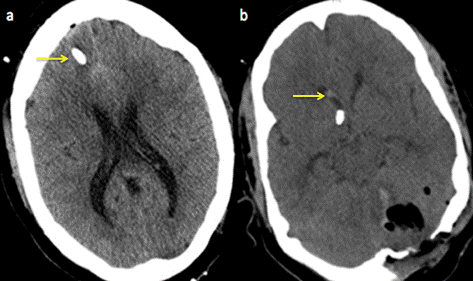
Figure 1. (a) Postoperative noncontrast cranial computed tomography demonstrates EVD placement (arrow) without associated ICH. (b) A small amount of IVH (arrow) is appreciated in the right frontal horn and the left cerebellar hematoma has been evacuated.
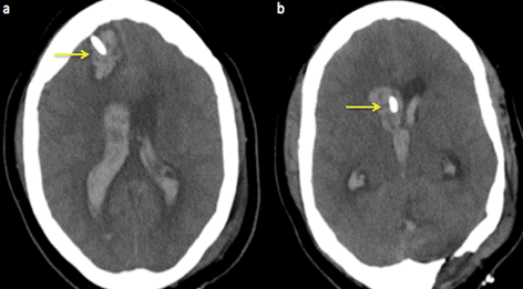
Figure 2. (a and b) Noncontrast cranial computed tomography demonstrates evidence of ICH along the EVD catheter extending into the ventricular system with obstructive hydrocephalus (arrows).
On postoperative day three, the patient experienced a sudden headache associated with spontaneous bloody cerebrospinal fluid (CSF) drainage from the EVD catheter, followed rapidly by acute neurologic deterioration. After intubation and initial resuscitation, an emergent bedside CT scan showed hemorrhage along the EVD tract with ventricular extension, casting of the ventricular system and obstructive hydrocephalus. (Fig 2, a and b) A contralateral EVD was placed, with minimal neurological improvement. CT angiography did not show any clear vascular abnormalities. A cerebral diagnostic angiogram revealed a 3.6 x 3.4 mm traumatic pseudoaneurysm arising from a distal anterior cerebral artery (ACA) branch that was in contact with the ventricular catheter. (Fig 3 a and b) Attempted endovascular obliteration of the pseudoanerysm was unsuccessful. Due to mass effect and increased intracranial pressure it was decided to take the patient to the operating room for clot evacuation and surgical pseudoaneurysm obliteration. Despite a long and complicated hospital course thereafter, her neurologic exam did not improve, and the patient subsequently expired.
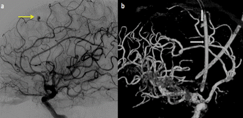
Figure 3. (a) Lateral digital substraction angiogram demonstrates a pseudoaneurysm in a distal branch of the ACA (arrow), (b) 3-D digital substraction angiography demonstrates the pseudoaneurysm along the location of the EVD catheter (arrows).
3. Discussion
In 1918 Dandy reported that, in experienced hands, ventricular puncture could be an uncomplicated procedure.4) Unfortunately, more experience and regular usage has demonstrated that EVD placement is associated with significant infectious and hemorrhagic risks with hemorrhagic complication rates ranging from 0-41%.5, 6) Two meta-analysis of this procedure have reported an overall hemorrhagic complication rate of 5.7-7%, with a significant rate of hemorrhage between 0.8%-1%.2, 3) The wide variation in hemorrhage rates may be associated with specific technical or anatomic factors, but could also be related to methodological differences. Variations in patient populations, coagulation profile thresholds prior to the procedure, routine post-procedure imaging studies and its timing, inclusion of hemorrhages caused by ventricular catheter removal, and operator technique/expertise may be important factors contributing to the broad range of results, as well as the retrospective nature of most studies.
Drain related hemorrhages (subdural, epidural, intracerebral and intraventricular-IVH) are all possible, most probably occur as the result of a small cortical or pial vessel injuries caused by the ventricular catheter or drill at the time of insertion. In most cases, they are not identified because they are asymptomatic and do not cause any clinical consequences, and therefore are excluded from published series. The few cases have been reported have rarely required surgical evacuation for mass effect.7-9)
We did not find any reported cases of ICH or IVH proven to be the consequence of a vessel injury leading to pseudoaneurysm formation and rebleeding following EVD. Thus, this phenomenon may be clinically quite rare. One possible explanation may be that in the majority of cases, the small hemorrhages related to vessel injury do not expand into a visible pseudoaneurysm, and the vessel heals without consequences. In the absence of clinical sequelae, they remain undetected, with rarely a need to obtain further diagnostic studies.
In a recently published series of complications related to EVD placement, 10) one of the patients developed a 3-mm pseudoaneurysm arising from a distal right ACA branch that was adjacent to the shunt tubing. Interestingly this pseudoaneurysm was not seen on angiography performed a month earlier and a CT scan after insertion of the EVD did not demonstrate evidence of hemorrhage. A possible explanation of the delay presentation may be related to formation of the aneurysm secondary to thinning of the vessel wall due to a chronic foreign body reaction secondary to the shunt tubing, as previously reported in a patient with traumatic aneurysm formation following ventriculoperitoneal shunt insertion. 11)
Pseudoaneurysms of the superficial temporal artery secondary to partial laceration of the vessel wall during tunneling of the ventricular catheter10, 12), and dural arteriovenous fistulas after injury of the middle meningeal artery during EVD placement have been previously reported.10, 13, 14) Schuette et al, 15) described a case of a pial arteriovenous fistula resulting from EVD placement. In this case a CT scan after drain placement demonstrated a hemorrhage at the EVD site. Interestingly, the fistula was appreciated on follow-up cerebral angiography 18 months after the EVD was initially inserted, and a previous 6 month follow-up angiogram did not reveal the lesion. This fistula was almost certainly the result of a prior clinically silent EVD related vessel injury that recanalized over time and became an arteriovenous fistula. To our knowledge, the case presented here is the first report of an ICH as a consequence of rupture of a pseudoaneurysm that developed soon after EVD placement.
In the case of our patient, a routine postoperative CT scan after cerebellar clot evacuation showed no ICH and a small asymptomatic IVH likely the result of bleeding as the EVD entered the ventricle (Fig 1 a and b). After later clinical deterioration caused by ICH and IVH, an emergent CT angiogram did not show any vascular abnormalities. A formal cerebral diagnostic angiogram, however, demonstrated the pseudoaneurysm arising from the distal ACA that appeared to be in contact with the EVD. (Fig 3b) While misplacement of EVD catheters particularly those traversing the interhemispheric fissure into the contralateral hemisphere can potentially increase the risk of vascular injury and potential pseudoaneurysm formation. In the case of our patient the EVD catheter remained ipsilateral along its course, and the tip of the catheter ended in the frontal horn. (Fig 1a and b).
4. Conclusion
Although percutaneous EVD is generally a safe and effective procedure, iatrogenic vascular trauma associated with this procedure may be more common than currently appreciated, and serious complications can arise. The presence of an expanding hematoma along the EVD tract in a patient with an uneventful insertion and with a normal coagulation profile may suggest the presence of vascular injury related to its placement. In such circumstances, early diagnosis and treatment may prevent the negative consequences of rebleeding related to pseudoaneurysm rupture. CT angiography may not be sufficient in the search of this lesion and cerebral angiography should be considered.
References
- Roitberg BZ, Khan N, Alp MS, Hersonskey T, Charbel FT, Ausman JI: Bedside external ventricular drain placement for the treatment of acute hydrocephalus. Br.J.Neurosurg. 15: 324-327, 2001.
- Binz DD, Toussaint LG,3rd, Friedman JA: Hemorrhagic complications of ventriculostomy placement: a meta-analysis. Neurocrit Care. 10: 253-256, 2009.
- Bauer DF, Razdan SN, Bartolucci AA, Markert JM: Meta-analysis of hemorrhagic complications from ventriculostomy placement by neurosurgeons. Neurosurgery 69: 255-260, 2011
- Dandy WE: Ventriculography Following the Injection of Air into the Cerebral Ventricles. Ann.Surg. 68: 5-11, 1918
- Gardner PA, Engh J, Atteberry D, Moossy JJ: Hemorrhage rates after external ventricular drain placement. J.Neurosurg. 110: 1021-1025, 2009.
- Khanna RK, Rosenblum ML, Rock JP, Malik GM: Prolonged external ventricular drainage with percutaneous long-tunnel ventriculostomies. J.Neurosurg. 83: 791-794, 1995.
- Kakarla UK, Kim LJ, Chang SW, Theodore N, Spetzler RF: Safety and accuracy of bedside external ventricular drain placement. Neurosurgery 63: ONS162-6; discussion ONS166-7, 2008.
- Saladino A, White JB, Wijdicks EF, Lanzino G: Malplacement of ventricular catheters by neurosurgeons: a single institution experience. Neurocrit Care. 10: 248-252, 2009.
- Maniker AH, Vaynman AY, Karimi RJ, Sabit AO, Holland B: Hemorrhagic complications of external ventricular drainage. Neurosurgery 59: ONS419-24; discussion ONS424-5, 2006.
- Kosty J, Pukenas B, Smith M, Storm PB, Zager E, Stiefel M, Leroux P, Hurst R: Iatrogenic Vascular Complications Associated with External Ventricular Drain Placement: A Report of Eight Cases and Review of the Literature. Neurosurgery DOI: 10.1227/NEU.0b013e318279e783, 2012.
- Jenkinson MD, Basu S, Broome JC, Eldridge PR, Buxton N: Traumatic cerebral aneurysm formation following ventriculoperitoneal shunt insertion. Childs Nerv.Syst. 22: 193-196, 2006.
- Angevine PD, Connolly ES,Jr: Pseudoaneurysms of the superficial temporal artery secondary to placement of external ventricular drainage catheters. Surg.Neurol. 58: 258-260, 2002.
- Field M, Branstetter BF,4th, Levy E, Yonas H, Jungreis CA: Dural arteriovenous fistula after ventriculostomy. Case illustration. J.Neurosurg. 97: 227, 2002.
- Meisel K, Yee A, Stout C, Kim W, Cooke D, Halbach V: Arteriovenous fistula after ventriculostomy in aneurysmal subarachnoid hemorrhage. Neurology 80: 2168, 2013.
- Schuette AJ, Blackburn SL, Barrow DL, Cawley CM: Pial arteriovenous fistula resulting from ventriculostomy. World Neurosurg. 77: 785.e1-785.e2, 2012.


