
American Journal of Clinical Neurology and Neurosurgery, Vol. 1, No. 2, September 2015 Publish Date: Aug. 13, 2015 Pages: 102-106
Impact of Preoperative Spinal Cord Signal Intensity and Symptom Duration on Surgical Outcome of the Patients with Cervical Spondylotic Myelopathy
Farzad Omidi-Kashani1, *, Mohamed Hosein Ebrahimzadeh2, Arezoo Naderimoghadam3, Hamid Amanzadeh4
1Orthopedic Research Center, Orthopedic Department, Imam Reza Hospital, Mashhad University of Medical Sciences, Mashhad, Iran
2Orthopedic Research Center, Orthopedic Department, Qaem Hospital, Mashhad University of Medical Sciences, Mashhad, Iran
3Student Research Committee, Faculty of Medicine, Imam Reza Hospital, Mashhad University of Medical Sciences, Mashhad, Iran
4Orthopedic Department, Imam Reza Hospital, Mashhad University of Medical Sciences, Mashhad, Iran
Abstract
Background: Cervical spondylotic Myelopathy (CSM) is one of the most common causes of cervical spinal cord dysfunction in old patents and in those cases with refractory complains, surgical decompression may become necessary. We aim to evaluate the impact of preoperative spinal cord signal change and symptom duration on surgical outcome of these patients. Material & Method: In this retrospective study, we reviewed 36 patients (25 male and 11 female) with CSM and mean age 55.7±13.2 who were surgically treated in our orthopedic department. Preoperative duration of myelopathic symptoms, disability status (according to Neck Disability Index; NDI), signal change inside the cervical spinal cord (on T2-weighted magnetic resonance imaging; T2 MRI), and surgical outcome (based on Odom’s criteria) were especially assessed. Statistical Package for the Social Sciences (SPSS) software version 16 was used for statistics and P value<0.05 was interpreted as significance. Results: Mean duration of preoperative symptoms and follow-up periods were 17.5±7.9 and 37.5±8 months, respectively. Spinal cord signal intensity in T2 MRI was normal in 21 (58.3%), light in 9 (25%), and tense in 6 (16.7%). Surgery could improve NDI scores from 27±9.3 preoperatively to 11.4±12 at the last follow-up visit. Excellent or good surgical outcome could achieve in 28 patients (77.8%). Among the various patients’ characteristics, only preoperative symptom duration and cord enhancement could show a significant correlation with patient’s disability improvement and surgical outcome. Conclusions: Outcome of surgery in CSM is satisfactory in most of the patients although, preoperative duration of myelopathic symptoms and spinal cord signal enhancement were associated with lower disability improvement and poorer surgical outcome.
Keywords
Spondylosis, Cervical Cord, Outcome Assessment, Magnetic Resonance Imaging
Received: July 18, 2015
Accepted: August 8, 2015
Published online: August 13, 2015
@ 2015 The Authors. Published by American Institute of Science. This Open Access article is under the CC BY-NC license. http://creativecommons.org/licenses/by-nc/4.0/
1. Introduction
As the cervical intervertebral disc degenerates, normal biomechanics of the cervical spine gradually changes and greater stress is applied on the surrounding structures and progressively a full blown cervical spondylosis may occur.1 Cervical spondylotic changes can lead to spinal stenosis including radiculopathy or myelopathy. Reduction in space available for the cord gradually induce white matter demyelination, gray matter neuronal loss, necrosis, cavitation and ultimately, white matter gliosis and diffuse gray matter necrosis may follow.2, 3 On the other hand, as motion and weight are greater in lower cervical spine, more degenerative changes are usually seen at C5-6 and C6-7 levels.4 This area of the cervical spine is also a watershed region in the cervical cord, with limited blood supply and consequently greater potential for spinal cord ischemia.5
Cervical spondylotic myelopathy (CSM) is one of the most common causes of cervical spinal cord dysfunction in old patents and in those cases with refractory complains or progressive neurologic deficit, surgical decompression may become necessary.6 There are many poor prognostic factors proposed for surgical outcome of the patients with CSM, but still debate exists about their definite roles. Old age at the time of surgery, loss of normal cervical lordotic curve, multi-segmental cord compression, and numerous underlying diseases are relatively known factors.7-10 In this study we aim to evaluate the impact of preoperative spinal cord signal change and symptom duration on surgical outcome of these particular patients.
2. Methods
Local institutional review board approval (registration number 920349) was first obtained and then we retrospectively began to gather the demographic, clinical, and paraclinical data from the medical records related to our surgically treated patients with CSM who had been treated from April 2010 to April 2013 in Orthopedic Department, Imam Reza Hospital, Mashhad, Iran.
2.1. Inclusion and Exclusion Criteria
Our inclusion criteria included all the patients with documented CSM based on both imaging and clinical characteristics, presence of significant neurologic deficit (like deep tendon reflex or motor disturbances), intractable complains longer than 6 months, and those patients signed the informed consents. Those myelopathic patients who were not followed-up for more than two years, could not stop smoking during the perioperative time, had a previous history of cervical spine surgery, those with significant associated diseases (like uncontrolled diabetes mellitus or hemorrhagic diathesis), or those with underlying etiologies other than degenerative disease (e.g. cancer, infection, or trauma) were excluded from the study. In this study, we did not care about the exact surgical technique or approach and our primary aim was efficacious neural decompression and spinal stabilization, if necessary.11
2.2. Patients Assessment
Preoperative symptom duration was assumed as the time between the beginning of clinical manifestation and surgical procedure. We evaluated patients’ disability with neck disability index (NDI) questionnaire.12, 13 In this study, NDI was expressed as its raw score (zero to fifty). This questionnaire was completed preoperatively and at the last follow-up visit.
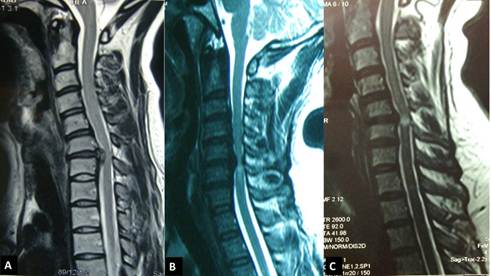
Fig. 1. Grading of cord enhancement based on increased signal intensity on T2 weighted MRI. A- Cord enhancement Grade 0, B- Cord enhancement Grade I, C- Cord enhancement Grade II. Note that signal intensity at the narrowest part of the cervical cord is identical with cerebrospinal fluid in this grade.
Signal change inside the cervical spinal cord (Fig. 1) was graded preoperatively based on amount of signal enhancement at the narrowest part of the cord on the sagittal T2 weighted magnetic resonance imaging (MRI) scans as grade 0 or none (without any signal enhancement in spinal cord), grade 1 or light (with mild increased signal intensity inside the spinal cord), and grade II or tense (with severe increased signal intensity or bright signal identical with cerebrospinal fluid).14 At the last follow-up visit, surgical outcome was assessed according to Odom’s criteria and expressed as excellent, good, fair, or poor based on amount of improvement in preoperative symptoms and complains.15
3. Statistical Analysis
Statistical Package for the Social Sciences (SPSS) software version 16 was used for statistical analysis, data management and documentation. P value<0.05 was interpreted as significance. We used Kolmogorov– Smiranov Test, Pearson correlation coefficient, Spearman rank correlation, independent T test, and Mann-Whitney test in this analysis.
4. Results
Due to lack of proper follow-up assessment, four patients were excluded from the study. After all inclusion and exclusion criteria were assumed into account, we ultimately studied 36 patients comprising 25 male (69.4%) and 11 female (30.6%). Mean age of the patients was 55.7±13.2 (range; 31-80 years). Mean duration of preoperative symptoms and follow-up periods were 17.5±7.9 (range; 5-31 months) and 37.5±8 (range; 25-60 months), respectively. More than half of the patients (52.7%) were followed-up for greater than three years. Preoperative imaging characteristics of our patients were depicted in Table 1. NDI in preoperative and postoperative era at the last follow-up visit and surgical outcome at this latest time were also shown in Table 2. Mean improvement in NDI during this follow-up period was 15.6±11.
Table 1. Preoperative imaging characteristics of our treated patients.
| Index | Number | Percentage (%) |
| Spinal cord signal change (T2 MRI): | ||
| Grade 0 | 21 | 58.3 |
| Grade I | 9 | 25 |
| Grade II | 6 | 16.7 |
| Cervical stenotic levels: | ||
| 1 level | 11 | 30.6 |
| 2 levels | 10 | 27.8 |
| 3 levels | 8 | 22.2 |
| 4 levels | 3 | 8.3 |
| 5 levels | 4 | 11.1 |
Table 2. Disability improvement and surgical outcome in the operated patients.
| Index | Preoperative | Postoperative at the most recent time |
| NDIà | 27±9.3 | 11.4±12 |
| Surgical outcome | ||
| Excellent | - | 24 (66.7%) |
| Good | - | 4 (11.1%) |
| Fair | - | 2 (5.6%) |
| Poor | - | 6 (16.7%) |
à NDI: Neck Disability Index
Relationships between variety of patients’ characteristics with disability improvement and surgical outcome were depicted in Table 3. As this table shows, the only parameters that significantly and reversely correlated with patient’s disability improvement and surgical outcome were preoperative symptom duration and the amount of cord enhancement.
Table 3. Relationship between patients’ characteristics and clinical results.
| Index | Disability improvement | Surgical outcome |
| (P value) | (P value) | |
| Sex | 0.531 | 0.924 |
| Age | 0.395 | 0.676 |
| Preoperative symptom duration | 0.013à (r: -0.41) | 0.025à (r: -0.21) |
| Number of stenotic levels | 0.921 | 0.964 |
| Follow-up duration | 0.298 | 0.179 |
| Cord enhancement (T2 MRI) | 0.012à (r: -0.25) | 0.006à (r: -0.18) |
à Significant statistically
5. Discussion
In this study, we tried to elucidate the impact of some preoperative characteristics on surgical outcome and disability improvement of the patients with CSM. Although the patient’s age, sex, number of stenotic levels, and amount of follow-up duration have no prognostic role, we could find that preoperative symptom duration and cord enhancement have a reverse relationship with disability improvement and outcome of the surgery.
A review on the literature reveals that numerous studies have been carried out on prognostic effect of spinal cord enhancement in the surgical outcome of CSM. Most of these studies like us, agreed with the poor prognostic effect of spinal cord signal change.9, 10, 16-20 Haddadian et al. in a prospective study evaluated clinical symptoms, signs, and imaging characteristics of 43 patients with CSM to find out their significance in surgical procedures. They found that atrophy of the hand muscles, preoperative spastic gait and cord atrophy as evident on MRI comprised poor prognostic factors in this decompressive surgery.16Similarly, Chatley and co-authors in 2009, published the results of their study on 64 cases with CSM who underwent surgery.17 They concluded that multisegmental cord enhancement on T2-weighted MRI was associated with lower functional recovery. There are fewer studies that suggest spinal cord signal change has no effect on the prognosis of the patients with CSM. Karpova et al. in a prospective study evaluated 65 patients with CSM.8 In contrary to our study, they quantified disability with modified Japanese Orthopaedic Association scale. Their results showed that younger age and higher preoperative disability scores were strongly associated with better recovery ratio. They could not find any relationship between spinal cord signal change or preoperative symptom duration and surgical outcome at the end of 12 months follow-up. This recent finding is completely contradicted with the results of the study we had carried out.
About the effectiveness of preoperative duration of myelopathic symptoms on the results of surgery, our findings are in line with the results of most other studies.8, 21, 22 Tetreault et al. in a systematic review of the literature found that preoperative severity and duration of myelopathic symptoms are the most predictive of surgical outcome.22 Similarly,Pumberger et al. reviewed and analyzed 284 patients with CSM and concluded that preoperative duration of myelopathic symptoms is a poor prognostic factor for surgical outcome but does not necessarily indicate higher severity of the disease.21 Although the study conducted by Karpova failed to show any relationship between the two.8
Our study had several significant flaws. We did not use any image analysis software to quantify the signal change of the spinal cord and just trusted our vision. Obviously, if we could carried out a prospective study on a larger number of patients using an appropriate computer software, the more reliable results would be achieved.
6. Conclusions
Outcome of surgery in CSM is satisfactory in most of the patients although, preoperative duration of myelopathic symptoms and spinal cord signal enhancement may be associated with lower disability improvement and poorer surgical outcome.
Acknowledgements
This study is funded by Student Research Committee, Faculty of Medicine, Mashhad University of Medical Sciences, Mashhad, Iran.
References
- Ferrara LA. The biomechanics of cervical spondylosis. Adv Orthop. 2012; 2012: 493605.
- Tracy JA, Bartleson JD. Cervical spondylotic myelopathy. Neurologist. 2010; 16(3): 176-87.
- Matz PG, Anderson PA, Holly LT, et al. The natural history of cervical spondylotic myelopathy. J Neurosurg Spine. 2009; 11(2): 104-11.
- Morishita Y, Hida S, Miyazaki M, et al. The effects of the degenerative changes in the functional spinal unit on the kinematics of the cervical spine. Spine. 2008; 33(6): E178-82.
- Hsu CY, Cheng CY, Lee JD, et al. Clinical features and outcomes of spinal cord infarction following vertebral artery dissection: a systematic review of the literature. Neurol Res. 2013; 35(7): 676-83.
- Sah S, Wang L, Dahal M, Acharya P, Dwivedi R. Surgical management of cervical spondylotic myelopathy. JNMA J Nepal Med Assoc. 2012; 52(188): 172-7.
- Furlan JC, Kalsi-Ryan S, Kailaya-Vasan A, Massicotte EM, Fehlings MG. Functional and clinical outcomes following surgical treatment in patients with cervical spondylotic myelopathy: a prospective study of 81 cases. J Neurosurg Spine 2011; 14(3): 348-55.
- Karpova A, Arun R, Davis AM, et al. Predictors of Surgical Outcome in Cervical Spondylotic Myelopathy. Spine 2013; 38(5): 392-400.
- Naderi S, Ozgen S, Pamir MN, Ozek MM, Erzen C. Cervical spondylotic myelopathy: surgical results and factors affecting prognosis. Neurosurgery 1998; 43(1): 43-9; discussion 49-50.
- Tetreault LA, Nouri A, Singh A, Fawcett M, Fehlings MG. Predictors of outcome in patients with cervical spondylotic myelopathy undergoing surgical treatment: a survey of members from AOSpine International. World Neurosurg. 2014; 81(3-4): 623-33.
- Mummaneni PV, Kaiser MG, Matz PG, et al. Cervical surgical techniques for the treatment of cervical spondylotic myelopathy. J Neurosurg Spine 2009; 11(2): 130-41.
- Vernon H, Mior S. The neck disability index: a study of reliability and validity. J Manipulative Physiol Ther 1991; 14(7): 409-15.
- Mousavi SJ, Parnianpour M, Montazeri A, et al. Translation and validation study of the Iranian versions of the neck disability index and the neck pain and disability scale. Spine 2007; 32(26): E825-31.
- Yukawa Y, Kato F, Yoshihara H, Yanase M, Ito K. MR T2 image classification in cervical compression myelopathy: predictor of surgical outcomes. Spine 2007; 32(15): 1675-8.
- Odom GL, Finney W, Woodhall B. Cervical disk lesions. J Am Med Assoc 1958; 166(1): 23-8.
- Haddadian K, Rezaei O, Sadeghi S, et al. Cervical spondylotic myelopathy: the pattern of neurologic deficits and improvement following anterior cervical decompression. Med J Islam Repub Iran 2005; 18(4): 331-5.
- Chatley A, Kumar R, Jain VK, Behari S, Sahu RN. Effect of spinal cord signal intensity changes on clinical outcome after surgery for cervical spondylotic myelopathy. J Neurosurg Spine 2009; 11(5): 562-7.
- Arvin B, Kalsi-Ryan S, Mercier D, Furlan JC, Massicotte EM, Fehlings MG. Preoperative magnetic resonance imaging is associated with baseline neurological status and can predict postoperative recovery in patients with cervical spondylotic myelopathy. Spine. 2013; 38(14): 1170-6.
- Tetreault LA, Dettori JR, Wilson JR, et al. Systematic review of magnetic resonance imaging characteristics that affect treatment decision making and predict clinical outcome in patients with cervical spondylotic myelopathy. Spine. 2013; 38(22 Suppl 1):S89-110.
- Vedantam A, Jonathan A, Rajshekhar V. Association of magnetic resonance imaging signal changes and outcome prediction after surgery for cervical spondylotic myelopathy. J Neurosurg Spine. 2011; 15(6): 660-6.
- Pumberger M, Froemel D, Aichmair A, et al. Clinical predictors of surgical outcome in cervical spondylotic myelopathy: an analysis of 248 patients. Bone Joint J. 2013; 95-B (7): 966-71.
- Tetreault LA, Karpova A, Fehlings MG. Predictors of outcome in patients with degenerative cervical spondylotic myelopathy undergoing surgical treatment: results of a systematic review. Eur Spine J. 2015; 24 Suppl 2: 236-51.


