International Journal of Plant Science and Ecology, Vol. 1, No. 5, October 2015 Publish Date: Aug. 13, 2015 Pages: 225-230
Synthesis of Silver Nanoparticles from Cymbopogon flexuosus Leaves Extract and Their Antibacterial Properties
Ashish Kumar Gupta, Deepak Ganjewala*
Amity Institute of Biotechnology, Amity University Uttar Pradesh (AUUP), Noida, India
Abstract
Here we report synthesis of silver nanoparticles using leaf extracts of four varieties of lemongrass (Cymbopogon flexuosus) namely krishna, neema, pragati and suvarna and their antibacterial activities against drug resistant bacteria Staphylococcus aureus, Pseudomonas aeruginosa and Acinatobacter bamunnii. Silver nanoparticles were synthesized by the bio-reduction of silver nitrate solution (1 mM) using water extracts of lemongrass leaves. Synthesis of silver nanoparticles was confirmed by the presence of an absorbance peak at 430-450 nm in UV-visible spectrum. Thus synthesized silver nanoparticles were analyzed by Dynamic Light Scattering (DLS) technique which revealed their z-average (nm) size 40-100 nm. The silver nanoparticles were tested for their antibacterial potential against drug resistant bacteria by agar well diffusion method. The results revealed that all the silver nanoparticles (4 mg/ml) tested exhibited strong antibacterial activities against S. aureus and P. aeruginosa with zone of inhibition ranged from 24-27 and 17-23 mm, respectively while less effective against A. bamunnii with zone of inhibition ranged 13-14 mm. In conclusion, lemongrass leaf extract can be used to synthesize silver nanoparticles of potential antibacterial activities against drug resistant bacteria.
Keywords
Antibacterial, Lemongrass, Silver Nanoparticles, Bio-Reduction, Drug Resistant Bacteria, Zone of Inhibition
Received: June 28, 2015
Accepted: July 18, 2015
Published online: August 13, 2015
@ 2015 The Authors. Published by American Institute of Science. This Open Access article is under the CC BY-NC license. http://creativecommons.org/licenses/by-nc/4.0/
Contents
1. Introduction 2. Materials and Methods 2.1. Plant Materials and Extraction 2.2. Preparation of Leaf Extracts 2.3. Synthesis of Silver Nanoparticles 2.4. Spectrophotometer Analysis 2.5. Dynamic Light Scattering (DLS) Analysis 2.6. Antibacterial Assay 3. Results 3.1. Synthesis of Silver Nanoparticles and Their Characterization 3.2. Antibacterial Activities 4. Discussion 5. Conclusion Acknowledgements
1. Introduction
Cymbopogon flexuosus (Steud.) is popularly known as lemongrass and locally as Cochin or Malabar grass is a tufted perennial grass (Weiss, 1997). It is grown in Kerala, Assam, Maharashtra and Uttar Pradesh. Apart from India, it is also cultivated in large scale in Brazil, Mexico, Dominica, Haiti, Madagascar, Indonesia and China (Ganjewala et al., 2008). Some elite cultivars of lemongrass are krishna, cauveri, pragati, chirharit, neema and suvarna which produce essential oil of wide applications in flavors, pharmaceutical and food industries. Lemongrass essential oil is mainly consisted of a monoterpene aldehyde citral (racemic mixture of two isomers called geranial and neral) which accounts for 75-85% of total monoterpene (Ganjewala and Gupta, 2013). Citral imparts characteristic lemon like aroma to the essential oil and is one of the most important constituent in flavors, fragrance and perfumery, for the synthesis of vitamin A and β-ionones (Dawson, 1994; Ganjewala et al., 2008; Ganjewala et al., 2012). Lemongrass leaf extract, essential oil and its constituents namely citral, geraniol, and geranyl acetate have been reported to possessed a number of bioactivities viz., antimicrobial, anti-inflammatory, anticancer, antioxidant, allelopathic, anthelmintic, and insect and mosquito repellent (Ganjewala et al., 2012; Ganjewala and Gupta, 2013). Lemongrass leaf extract can also be used for the synthesis of nanoparticles of silver and gold and tested for bactericidal effects against pathogenic bacteria. Shankar et al., (2004) have used lemongrass extract for the synthesis of gold nanoparticles by bio-reduction method which is also the first report on use of lemongrass extract to synthesize nanoparticles. Fierascu et al., (2010) have reported synthesis of gold nanoparticles from lemongrass natural extract and its use as plasmonic material. Silver nanoparticles synthesized from lemongrass extracts demonstrated antimicrobial activity against drug resistant bacteria (Masurkar et al., 2011). Nanoparticles of CeO2 synthesized using lemongrass extract are reported to exhibit optical properties (Maensiri et al., 2014).
Nanoparticles owing to their characteristic properties of size and structural similarity to biological molecules may have tremendous biomedical applications. Silver nanoparticles owing to their powerful bioactivities against bacteria, fungi, protozoa, and viruses have been considered most promising antimicrobial agents among others (Masurkar et al., 2011; Sharma et al., 2009). Nanoparticles can be synthesized by three methods: 1. chemical method such as reduction (Balantrapu et al., 2009; Tripathi et al., 2010), 2. Electro-chemical method (Rodriguez-Sanchez et al., 2000) and 3. bio-reduction method also called green synthesis (Begum et al., 2013). However, the bio-reduction method is most widely used method as it is safer, cost effective and sustainable compared to other methods (Gardea-Torresdey et al., 2002). A number of studies have reported the green synthesis of nanoparticles from plant extracts of Allium cepa (Saxena et al., 2010), Gliricidia sepium (Raut et al., 2009), Pongamia pinnata (L), Azadirachta indica (Shankar et al., 2004), Carica papaya (Mude et al., 2009). The green synthesis of nanoparticles using plant extracts is a rapidly growing area of the nano-biotechnology which attracted the attention of researchers from all over. In view of ever increasing significance of nano-biotehcnology in human life, the present study was undertaken with an objective to synthesize silver nanoparticle from lemongrass leaf extract of four cultivars by bio-reduction method and to test their antibacterial activities against the drug resistant bacteria or hospital isolates.
2. Materials and Methods
2.1. Plant Materials and Extraction
Lemongrass (Cymbopogon flexuosus Steud.) cultivars krishna, neema, pragati and suvrana plants (Figure 1) were grown in the Organic Farm House, Amity University Uttar Pradesh, India. Plants at 50 DAE (days after leaf emergence) when they are full matured were harvested for use in the study to prepare leaf extract.
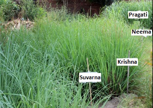
Figure 1. A view of lemongrass (C. flexuosus) cultivars: krishna, neema, pragati and suvrana.
2.2. Preparation of Leaf Extracts
Full grown plants of lemongrass cultivars were harvested from the field and leaves were separated and cut into small pieces. Cut leaves (10 g) were transferred into a beaker containing 50 ml of deionizer water and boiled for one hour at 80ºC in water bath to prepare an extract. Thus prepared extract was allowed to cool and filtered through Whatman paper No. 1 into a clean dry beaker. The transparent extract (filtrate) collected in the beaker was used to synthesize silver nanoparticles.
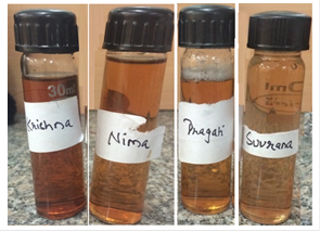
Figure 2. Photographs depicting synthesis of silver nanoparticles after 24 h of incubation of reaction mixture containing 1 mM silver nitrate and lemongrass extract (9:1 v/v).
2.3. Synthesis of Silver Nanoparticles
Synthesis of silver nanoparticles was achieved according to previously published reports (Masurkar et al., 2011; Logeswari et al., 2012). Freshly prepared aqueous solution (90 ml) of 1 mM AgNO3 and lemongrass leaf extract (10 ml) were mixed in a 150 ml conical flask and incubated for 24 h at room temperature. Periodically, flasks were observed for change in color of the reaction mixture from pale yellow to light-dark brown. The solution was further analysed by UV-visible spectroscopy.
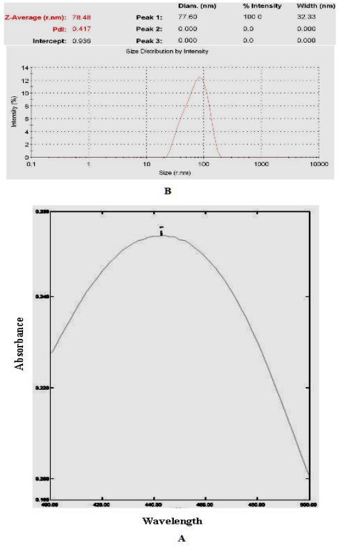
Figure 3. Ultraviolet-visible absorption (A) and DLS spectra (B) of silver nanoparticles synthesized from lemongrass leaf extract.
2.4. Spectrophotometer Analysis
The light brown solutions of silver nitrate were analyzed by UV-visible spectroscopy (UV-1601 pc Shimadzu) to confirm the bio-reduction of silver ions (Ag2+) into metallic silver nanoparticles by lemongrass leaf extract. The presence of a peak between 430-450 nm in UV-visible spectrum confirms the synthesis of silver nanoparticles.
2.5. Dynamic Light Scattering (DLS) Analysis
The colloidal solutions of silver nanoparticles were subjected to DLS analysis for determination of size of the nanoparticles at nano scale (Raliya and Tarafdar, 2012).
2.6. Antibacterial Assay
Antibacterial activities of the synthesized silver nanoparticles were tested against three drug resistant bacteria namely Acinetobacter bamunni, Pseudomonas aeruginosa, and Staphylococcus aureus by agar well diffusion method (Kakarla and Ganjewala, 2009). The drug resistant bacteria were obtained from Microbiology Department, Apollo Indraprashtha Hospitals, New Delhi, India and maintained in nutrient agar media (NAM) at 4 °C and 50 % glycerol stock at -20°C. Agar culture plates were prepared by pouring 20 ml of pre-sterilized nutrient agar (HiMedia, Mumbai, India) in to Petri dishes. The plates were inoculated using 1 mL (1.0×10⁷cfu) of 24 h bacterial cultures by spread plating method. Then wells of 6 mm diameter were made in the inoculated agar plates with the help of a sterile borer. Solutions of the silver nanoparticles (50 μL, 75 μL, and 100 μL) were introduced into separate wells with the help of a micropipette. Standard antibiotics such as ampicillin (10µg/disc), gentamicin (10µg/disc), rifampicin (5µg/ml), tetracycline (10µg/ml) and vancomycin (30μg/ml) were used as positive control and silver nitrate solution as the negative control. The agar plates were incubated at 37°C for 24 h. After incubation, the plates were observed and diameters of zone of inhibition (mm) were measured. The antibacterial assay was performed in triplicates.
3. Results
3.1. Synthesis of Silver Nanoparticles and Their Characterization
Silver nanoparticles (AgNPs) were synthesized from leaf extracts of four lemongrass cultivars namely krishna, neema, pragati and suvarna by bio-reduction method. Synthesis of the AgNPs was confirmed first by visual observation of the change in the color of AgNO3 solution as a result of bio-reduction of Ag+by plant extract. The Figure 1 shows the change of color from pale yellow to dark brown indicating the bio-reduction of Ag+ into AgNPs. Bio-reduction of Ag+ into metallic silver by leaf extracts begins after 8-10 minutes of incubation. In the control no change in the color was observed.
Analyses of the brown colloidal solutions of silver nanoparticles by UV-vis spectroscopy revealed the presence of a peak between 430-450 nm in absorption spectra (Fig. 3A). The z-average (nm) size of the AgNPs determined by DLS technique was in the range of 40-100 nm (Fig. 3B).
3.2. Antibacterial Activities
The results of antibacterial assay presented in the Table 1 revealed that synthesized AgNPs showed strong antibacterial activities against the drug resistant S. aureus and P. aeruginosa while weak activity against A. bamunni. Antibacterial activities increase with increase in the concentration of synthesized AgNPs. All the AgNPs at 150 µL concentration exhibited highest antibacterial activities against S. aureus and P. aeruginosa. The diameter of zone of inhibition determined for S. aureus was ranged from 24-27 mm while for P. aeruginosa 17-23 mm. The diameter of zone of inhibition for A. bamunnii measured was 13-14 mm. Antibacterial activities of the synthesized AgNPs were comparable to the standard antibiotics viz., ampicillin, vancomycin, gentamicin, rifampicin and tetracycline (Table 1). Among all gentamicin was found to be most effective antibiotics followed by vancomycin and tetracylin against drug resistant bacteria used. Two antibiotics ampicillin and rifampicin did not show any activity against the bacteria used (Table 1).
Table 1. Antibacterial activities of colloidal silver nanoparticles against the drug resistant bacteria.
| Synthesized AgNPs | Concentrations | Zone of inhibition (mm) | ||
| A. bamunnii | P. aeruginosa | S.aureus | ||
| Krishna | 50 µL | 11 | 18 | 20 |
| 100 µL | 13 | 20 | 22 | |
| 150 µL | 14 | 22 | 24 | |
| Neema | 50 µL | 12 | 20 | 23 |
| 100 µL | 13 | 19 | 25 | |
| 150 µL | 14 | 17 | 27 | |
| Pragati | 50 µL | 11 | 18 | 23 |
| 100 µL | 13 | 20 | 25 | |
| 150 µL | 14 | 23 | 26 | |
| Suvarna | 50 µL | 10 | 19 | 21 |
| 100 µL | 11 | 21 | 24 | |
| 150 µL | 13 | 22 | 27 | |
| Antibiotics | ||||
| Ampicillin | 10mcg/disc | Nil | Nil | Nil |
| Vancomycin | 30mcg/disc | 11 | 13 | 23 |
| Gentamicin | 10mcg/disc | 19 | 20 | 33 |
| Rifampicin | 5mcg/disc | Nil | Nil | Nil |
| Tetracycline | 10mcg/disc | 11 | 17 | 11 |
4. Discussion
Tremendous progress is being witnessed in global attempts to discover newer antimicrobials from plants against the bacteria rapidly evolving resistant mechanisms against available drugs. Nanoparticles of gold, silver, iron and others metals synthesized using extracts of a variety plants have shown remarkable antimicrobial potential thus paved the way for development of new powerful antibiotics against the multi drug resistant bacteria. Nanoparticles have offered promises from early diagnosis of diseases including those caused by emerging multidrug-resistant pathogens to their management. A number of reports showed the antibacterial potential of the silver nanoparticles synthesized from plant extracts against myriad of microorganisms. The present report describes the synthesis of AgNPs using leaf extracts of four lemongrass cultivars which display significant antibacterial activity against drug resistant bacteria. Synthesis of silver nanoparticles using lemongrass leaf extracts by bio-reduction method was in accordance to a number of previously published reports. The bio-reduction of Ag+ into metallic silver Ag is suggested to be catalyzed by different bio-molecules such as reducing sugars, proteins, terpenoids, and phenolic compounds present in the plant extracts (Cooper and Kavanagh, 1972). In the present study, the colorless solution of AgNO3: leaf extract (9:1 v/v) initially changed to light yellow and finally to a dark-brown color indicated the synthesis of AgNPs. The change in color was visible after 8-10 minutes of incubation. The synthesis of AgNPs their size and stability is influenced by temperature and pH (Raut et al., 2011; Shankar et al., 2004). Increase in temperature accelerates the synthesis of AgNPs. Here, the synthesis of AgPNs using lemongrass leaf extracts was carried out at room temperature similar to a previous report (Masurkar et al., 2011).
The formation and stability of synthesized AgNPs were initially tested by UV-vis spectroscopic analysis (Fig. 3A). The λmax was in the range of 430-450 nm which confirmed the synthesis of AgNPs. The appearance of a peak at 430-450 nm is due to AgNPs surface Plasmon absorbance (Akanna et al., 2010). The UV-Vis characteristics of the synthesized AgNPs are consistent with previously published reports (Masurkar et al., 2011; Mondal et al., 2011). Appearance of additional absorption peaks may be due to the presence of many participating organic compounds that can interact to reduce the silver ions. The z-average (nm) size of synthesized AgNPs as determined by the DLS technique was in the range of 40-100 nm (Fig. 3B). The DLS technique determines the size of nanoparticles by measuring the random changes in the intensity of light scattered from the suspension or solution. Silver nanoparticles solution resulted in Brownian motion of the particles and displayed the z-average size of all the particles in a solution; also it shows the peak and diameter of highest intensity particle present in the solution.
The synthesized AgNPs have demonstrated strong antibacterial activities against drug resistant bacteria S. aureus and P. aeruginosa (Table 1). Antibacterial activities of AgNPs were comparable to the standard antibiotics used. The antibacterial potential of the AgNPs reported here are similar to antibacterial potential of silver nanoparticles synthesized using C. citratus leaf extract (Masurkar et al., 2011). Antibacterial properties of the AgNPs may be due to their interactions with the cell wall of bacteria which results in the pore formation in cell walls where the AgNPs get deposited that causes change in permeability of the cell membrane (Grover et al., 2011; Cooper and Kavanagh, 1972). Also, AgNPs affects the proteins in the cytoplasm of the bacterial cells which lead to re-regulation in the functional cells and the DNA replication which will disrupt the replication mechanism leading to killing of the bacteria (Sondi et al., 2004).
5. Conclusion
The silver nanoparticles synthesized from lemongrass leaf extracts by bio-reduction method have exhibited all the characteristics features of the NPs. Most importantly, they demonstrated strong antibacterial activities against drug resistant hospital isolates of S. aureus, P. auriginosa. At present further studies to characterize the AgNPs by SEM and TEM and elucidation of mechanism of action of AgNPs are under progress. These studies would be certainly useful for development of AgNPs as an effective antimicrobial agent against the drug resistant microorganisms.
Acknowledgements
Corresponding author of this article is grateful to Dr. Ashok Kumar Chauhan, Founder President and Mr. Atul Chauhan, Chancellor, Amity University, Uttar Pradesh, Noida, India for providing necessary support and facilities.
References
- Akanna S., Prasad K.V., Elumalai E. and Savithramma N., 2010. Production of biogenic silver nanoparticles using Boswellia ovalifoliolata stem bark. Digest. Journal of Nanomaterial and Biostructures, 5:369-372
- Balantrapu K. and Goia D.V., 2009. Silver nanoparticles for printable electronics and biological applications. Journal of Material Research, 24:2828-2836
- Begum N.A., Mondal S., Basu S., Laskar R.A. and Mandal D., 2009. Biogenic synthesis of Au and Ag nanoparticles using aqueous solutions of black tea leaf extracts. Colloids and Surfaces B: Biointerfaces, 71:113-118
- Cooper RE. and Kavanagh F., 1972. Analytical microbiology. FW Kavanageh (Ed.), 1, 2.
- Dawson F.A., 1995. The amazing terpenes. Naval Stores Review, 104: 6-12
- Fierascu R.C., Dumitriu I. and Ion R., 2010.Plasmonic materials obtained in natural extract. Romanian Journal of Physics, 55:758-763
- Ganjewala D. and Gupta A.K., 2013. Lemongrass (Cymbopogon flexuosus Steud.) wats essential oil: overview and biological activities. Recent Progress in Medicinal and Aromatic Plants, 37:235-271
- Ganjewala D., Gupta A.K. and Muhury R., 2012. An update on bioactive potential of a monoterpene aldehyde citral. Journal ofBiologically Active Products from Nature, 2:186-199
- Ganjewala D., Kumari A. and Khan K.H., 2008. Ontogenic and developmental changes in essential oil content and compositions in Cymbopogon flexuosus cultivars. Recent Advance in Biotechnology, Excel India Publishers, New Delhi, pp. 82-92
- Gardea-Torresdey J.L., Parsons J.G., Gomez E., Peralta-Videa J., Troiani H.E., Santiago P. and Yacaman M.J., 2002.Formation and growth of Au nanoparticles inside live alfalfa plants. Nanoscience Letters, 2:397-401
- Grover V.A., Hu J., Engates K.E. and Shipley H.J., 2012. Adsorption and desorption of bivalent metals to hematite nanoparticles. Environmental Toxicology and Chemistry, 31:86-92
- Kakarla S. and Ganjewala D., 2009.Antimicrobial activity of essential oils of four lemongrass (Cymbopogon flexuosus Steud) varieties. Medicinal and Aromatic Plants Science Biotechnology, 3:107-109
- Logeswari P., Silambarasan S. and Abraham J., 2012. Synthesis of silver nanoparticles using plants extract and analysis of their antimicrobial property. Journal of Saudi Chemical Society, 19:311-317
- Maensiri S., Labuayai S., Laokul P., Klinkaewnarong J. and Swatsitang E., 2014. Structure and optical properties of CeO2 nanoparticles prepared by using lemongrass plant extract solution. Japanese Journal of Applied Physics, 53:06JG14
- Masurkar S.A., Chaudhari P.R., Shidore V.B. and Kamble S.P., 2011.Rapid biosynthesis of silver nanoparticles using Cymbopogan citratus (lemongrass) and its antimicrobial activity. Nano-Microbiology Letter, 3:189-194
- Mondal A.K., Mondal S., Samanta S. and Mallick S., 2011. Synthesis of ecofriendly silver nanoparticles from plant latex used as an important taxonomic tool for phylogenetic inter-relationship. Advances in Bioresearch, 2:122-133
- Mude N., Ingle A., Gade A. and Rai M., 2009. Synthesis of silver nanoparticles using callus extract of Carica papaya- a first report. Journal of Plant Biochemistry and Biotechnology, 18: 83-86
- Raliya R. and Tarafdar J.C., 2012. Novel approach for silver nanoparticle synthesis using Aspergillus terreus CZR-1: mechanism perspective. Journal of. Bionanoscience, 6:12-16
- Rodriguez-Sanchez L., Blanco M.C. and Lopez-Quintela M.A., 2000.Electrochemical synthesis of silver nanoparticles. The Journal of Physical Chemistry: B, 104:9683-9688
- Rout R.W., Lakkakula J.R. Kolekar N.S., Mendhulkar V.D. and Kashid S.B., 2009. Phytosynthesis of silver nanoparticle using Gliricidia sepium (Jacq.). Current Nanoscience, 5:117-122
- Saxena A., Tripathi R.M. and Singh R.P., 2010. Biological synthesis of silver nanoparticles by using onion (Allium cepa) extract and their antibacterial activity. Digest Journal of Nanomaterial and Biostructure, 5:427-432
- Shankar S.S., Rai A., Ankamwar B., Singh A., Ahmad A. and Sastry M., 2004. Biological synthesis of triangular gold nanoprisms. Nature Materials, 3:482-488
- Sharma V.K., Yngard R.A. and Lin Y., 2009. Silver nanoparticles: green synthesis and their antimicrobial activities. Advances in Colloid and Interface Science, 145:83-96
- Tripathi A., Chandrasekaran N., Raichur A.M. and Mukherjee A., 2009. Antibacterial applications of silver nanoparticles synthesized by aqueous extract of Azadirachta indica (Neem) leaves. Journal of Biomedical Nanotechnology, 5:93-98.
- Weiss E.A., 1997. Essential oil crops: Wallingford, UK, CAB International. pp. 59-137.


