International Journal of Plant Science and Ecology, Vol. 1, No. 5, October 2015 Publish Date: Aug. 10, 2015 Pages: 218-224
The Effect of Ethanolic Extract of Basella alba L. Leaves on Lead Induced Hepatotoxicity in Male Albino Wistar Rats (Rattus norvegicus)
Olubayode Bamidele*, Temitope Ibiere Adebiyi, Lawrence Dayo Babatunde, Dennis Seyi Arokoyo
Department of Physiology, Faculty of Basic Medical Sciences, College of Health Sciences, Bowen University, Iwo, Osun State, Nigeria
Abstract
Basella alba L. is claimed to be used as an antidote for poisons but has not been scientific adequately explored. This work investigated the effect of ethanolic extract of Basella alba on lead- induced hepatotoxicity in male rats. 24 male albino Wistar rats were randomly divided into 4 groups. Group 1 was given orally 10ml/kg normal saline while Group 2 received 50mg/kg of Lead Acetate for 4 weeks. Group 3 received 50mg/kg of Lead Acetate for first week and 250mg/kg Basella alba for 3 weeks. Group 4 received 250mg/kg of Basella alba for first week and 50mg/kg of Lead Acetate for the next 3 weeks. Then, levels of the liver enzymes in the serum were measured. The results showed that the levels of alkaline phosphatase, Aspartate aminotransferase and Alanine aminotransferase were significantly reduced while Glutathione was significantly increased in Basella alba treated Groups compared to Lead Control Group at (p < 0.05). The data suggests that Basella alba reduces lead toxicity.
Keywords
Basella alba, Lead, Hepatotoxicity, Liver Enzymes, Albino Rat
Received: June 25, 2015
Accepted: July 17, 2015
Published online: August 9, 2015
@ 2015 The Authors. Published by American Institute of Science. This Open Access article is under the CC BY-NC license. http://creativecommons.org/licenses/by-nc/4.0/
Contents
1. Introduction 2. Materials and Methods 2.1. Plant Materials 2.2. Experimental Animals 2.3. Experimental Design 2.4. Tissue Analysis of Lead Metal 2.5. Serum Analysis 2.6. Alanine Aminotransferase (ALT) and Aspartate Aminotransferase (AST) 2.7. Alkaline Phosphatase (ALP) 2.8. Glutathione (GSH) 2.9. Body Weight Measurement 2.10. Statistical Analysis 3. Results 3.1. Body Weight 3.2. Effect of Lead and Basella Alba on Body Weight 3.3. Effect of Lead and Basella Alba on Some Hepatic Biochemical Parameters 3.4. Alkaline Phosphatase 3.5. Aspartate Aminotransferase 3.6. Alanine Aminotransferase 3.7. Glutathione (GSH) 3.8. Lead Levels in the Experimental Male Albino Rats 4. Discussion 5. Conclusion
1. Introduction
Lead is a chemical element in the carbon group with symbol Pb (which is derived from a Latin word Plumbum) and atomic number 82. Lead which is a soft, grey-blue heavy metal is a common cause of poisoning in domestic animals throughout the world (Khan et al, 2008). Lead is a poisonous metal, which exist in both organic (Tetraethyl lead) and inorganic (lead acetate, lead chloride) forms in the environment (Shalan et al, 2005). It has been used in medicines, paintings, pipes, ammunition and in more recent times in alloys for welding storage materials for chemical reagents (Garaza et al, 2006).
Heavy metal pollution is one of the most important environmental problems today. Modern industries are to a large extent, responsible for contamination of the environment. Nriagu and Pacyna (1988) reported that industrial wastes contain various types of toxic chemicals. Three kinds of toxic heavy metals are of concern and these include lead. Heavy metals are dangerous because they tend to bioaccumulate.
Lead is a very strong poison. When a person swallows a lead object or breathes in lead dust, some of the poison can stay in the body and cause serious health problems (Woolf, 2007). Lead spreads so widely throughout the environment, it can be found in everyone’s body today. The levels found today in most people are orders of magnitude greater than that of ancient times (Flegal, 1995). These levels are within an order of magnitude of levels that have resulted in adverse health effects (Budd et al, 1998). The liver, kidneys, and brain are considered to be the target organs for the toxic effects of lead (Sharma and Street, 1980; Jackie et al, 2011). Toxicity of lead is mainly attributed to the induction of oxidative stress by elevation of reactive oxygen species (ROS) such as superoxide radicals, hydrogen peroxide and hydroxyl radicals and lipid peroxides, therefore, increased interest among phytotherapy investigators to use medicinal plants with antioxidant activity for protection against metal, especially lead, toxicity has been noted (Sharma and Street, 1980; Xu et al, 2005). Lead induced hepatotoxicity was reported to be associated with the impairments of liver structure and function (Aziz et al, 2012).
Natural products have been used for combating human diseases for thousands of years, since they exhibit a wide range of biological properties that can be exploited for medical application (Newman, 2003). Of the world population, about three quarter relies on plants and their extracts for their healthcare (Jachak and Saklani, 2007). Plants provide the possibility of an alternative strategy in exploration for new drugs (Abalaka et al, 2011). Infectious diseases, which account for the significant proportion of the health problems, are most often catered for by this system of medicine (Yasmin et al, 2009). Herbal drug analyses the part or parts of a plant used for the preparation of herbal and traditional medicines (for examples: leaves, flowers, seeds, roots, barks, stems, etc.) (Kayode and Kayode, 2011). Plants constitute many biologically active compounds that possess ability and criteria for development as medicinal agents (Abalaka and Oyewole, 2011).
Medicinal plants play a prominent role in the new era of medicine. Numerous medicinal plants and their formulations are used for various disorders in ethno medical practices as well as in the traditional system of India (Rodda et al, 2012). Among these plants is Basella alba L, a fast growing vegetable known to possess ant-inflammatory and antioxidant properties. Its composition includes proteins, fat, carbohydrates, fiber, ash, calcium, vitamins, thiamine, riboflavin, niacin. Traditionally it is used as an antidote, aperient, astringent, demulcent, diuretic, febrifuge, laxative, rubefacient and the juice of its fruits is used as a dye (Grubben and Denton, 2004). Basella alba L. contains basellasaponins found in the fresh aerial parts of the plant and it has been implicated to have antidote properties (Shi et al, 2004).
A research study by Olaniyi et al, in 2013 showed that certain bioactive agents with antioxidant and anti-lipid peroxidation properties are present in the Basella alba L. leaves extracts. This study gives scientific support for its wide consumption as leafy vegetable in western Nigeria and its exploration as a template in drug discovery and design especially for the management and prevention of diseases associated with oxidative damage, tissue degenerations and aging. In view of this, this work was carried out to investigate the effect of ethanolic extract of Basella alba L. leaves on lead induced hepatotoxicity in male albino wistar rats (rattus norvegicus).
2. Materials and Methods
2.1. Plant Materials
The basella alba plants were found at various humid locations in Iwo and Osogbo environments, Nigeria and their leaves were obtained. The plants were identified and authenticated by Mr Donatus Esimekhuai of the Department of Botany, University of Ibadan (UIH- 22391). The leaves were shade dried for four weeks after which they were ground into fine powder. Ethanol was used as a solvent for the extraction of the plant material by using Soxhlet apparatus. 100 grammes of powdered leaves were extracted in 1000ml of ethanol for three hours. The extract obtained was dried to a constant weight (20g) using a water bath at 60oC.
2.2. Experimental Animals
A total of 24 male Wistar albino rats with weight range of 180-220g purchased from Olu Research Farm, Sango, Oyo state, Nigeria were used in this study. The animals were kept in cages under standard conditions (temperature, 25±2°c, 12 hours light and 12 hours dark cycle) in the animal house of the Physiology Department, Faculty of Basic medical science, Bowen University, Iwo, Osun State, Nigeria. All animals were fed with commercially formulated rat feed and water ad libitum. After randomization into various groups, the rats were acclimatized for a period of 2 weeks in the environment before the initiation of the experiment. Their cages were cleaned of waste daily. All procedures involving the use of animals in this study complied with the guiding principles for research involving animals as recommended by the declaration of Helsinki and the Guiding principles in the care and use of animals (World Medical Association, 2002).
2.3. Experimental Design
Twenty four (24) Wistar strain albino rats were distributed into four groups of six animals per group.
Group 1: the control group received 1ml of normal saline by oral gavage.
Group 2: the lead control group received lead acetate daily for 4 weeks by oral gavage.
Group 3: the post-treatment group received lead acetate daily for the 1st week and was given basella alba from the 2nd week till the 4th week of the experiment. Both substances were administered by oral gavage.
Group 4: the pre-treatment group received basella alba for the 1st week and lead acetate from the 2nd week till the 4th week of the experiment. Both substances were administered by oral gavage.
After administration of last dose, the animals were given a rest overnight. They were anaesthetized with chloroform and blood samples were collected by cardiac puncture into ethylenediaminetetraacetic acid (EDTA) bottles. The livers of the rats were collected and rinsed and used for tissue analysis.
2.4. Tissue Analysis of Lead Metal
The liver tissue was ground using a ceramic mortar and pestle. The ground tissue was filled in plain serum bottle till it reached the 1cm mark. The plain serum bottle was filled with 2M H2SO4 till it got to the 4cm mark (Cretacci and Parsons, 2010). It was shaken and centrifuged for 20 minutes at 400 revolutions per minute. After centrifuging the clear fluid was decanted and analyzed for lead concentrations using the Flame Atomic Absorption Spectrophotometer (AAS) (Medham, 2000). Values were expressed in parts per million (ppm).
2.5. Serum Analysis
The serum of the blood collected by cardiac puncture into EDTA bottles was used for analysis of alkaline phosphatase (ALP), alanine aminotransferase (ALT), aspartate aminotransferase (AST) enzymes and glutathione (GSH).
2.6. Alanine Aminotransferase (ALT) and Aspartate Aminotransferase (AST)
Activities of ALT and AST were assayed by the method of Reitman and Frankel (1957). 0.1ml of serum was added to 0.5ml of phosphate buffer which contains L-alanine and α-oxoglutarate (for ALT) and L-aspartate and α-oxoglutarate (for AST) and it was incubated for 30 minutes at 37⁰C. The result was mixed with 0.5ml of 2,4-dinitrophenylhydrazine and allowed to stand for exactly 20 minutes at 20-25⁰C. Then 5.0ml of 0.4mol/l sodium hydroxide was added and left for 5mins. The absorbance was read against the sample blank at 546nm.
The sample blank solution: 0.5ml phosphate buffer was incubated at 37⁰C for 30 minutes. Then 0.5ml of 2,4-dinitrophenylhydrazine + 0.1ml of serum was added and allowed to stand for 20 minutes at 20-25⁰C then 5.0ml of sodium hydroxide was added. Activities were expressed as U/L.
2.7. Alkaline Phosphatase (ALP)
ALP was assayed according to the Rec. GSCC method (1972). 0.02ml of serum was mixed with 1.0ml of the reagent and incubated for 40 minutes at 37⁰C. The reagent is made up of 0.5mmol/l diethanolamine buffer (MgCl2) and 10mmol/l of p-nitrophenylphosphate. It was read at a wavelength of 405nm initially then re-read after 1,2 and 3 minutes. Activities were expressed as U/L.
2.8. Glutathione (GSH)
GSH was assayed according to the method of Ellman (1959). 0.2ml of sample is added to 1.8ml of distilled water. 3ml of the precipitating solution was added and allowed to stand for 5 minutes. (The precipitating solution is made up of 1.67g glacial metaphosphoric acid, 0.2 disodium EDTA and 30g sodium chloride in 100ml of distilled water). After standing for 5 minutes the solution was filtered and 2ml of the filtrate was added to 8.0ml of phosphate (0.3M disodium orthophosphate solution in distilled water). Then 5ml of the resulting solution was added to 1ml of DTNB (5.5 dithiobis-2-nitrobenzoic acid) reagent. The DTNB reagent was prepared by adding 40mg of DTNB in 100ml of 1% sodium citrate. It was measured at 412nm against blank. Activities were expressed as U/L.
2.9. Body Weight Measurement
The body weights of the rats were determined before the start of the experiment. During the experiment the rats were weighed at the end of each week. The measurements were taken using a sensitive weighing balance.
2.10. Statistical Analysis
Data are expressed as the mean ± SEM. The data were analyzed by analysis of variance (ANOVA) followed by Duncan test using the Statistical Package for the Social Sciences (S.P.S.S). The level of significance was set as p<0.05 and p<0.01.
3. Results
3.1. Body Weight
The body weights of the rats (measured in grammes) are expressed in mean ± standard error of mean. The rats were weighed before the experiment (week 0) and at the end of each week during the experiment (weeks 1-4). This is shown in the table 1.
3.2. Effect of Lead and Basella Alba on Body Weight
The body weight of rats treated with lead and Basella alba is represented in table 1. When the body weight of rats in group 3 (Post-treatment Group) was compared with the Healthy Control Group (group1) there was an increase in body weight that is not statistically significant (p>0.05). There was a slight increase in body weight in the first week and a decline from the second to the fourth in Group 4 (Pre-treatment Group) compared with the healthy control group (Group 1) but not statistically significant. When Lead Acetate Control Group (Group 2) was compared with the Healthy Control Group (Group 1) there was progressive weight loss (p<0.05) which was significant only in the fourth week.
Table 1. Effect of lead acetate and basella alba on body weight in male albino rats (n=6).
| GROUPS | BODY WEIGHT(g) WEEK 0 | BODY WEIGHT(g) WEEK 1 | BODY WEIGHT(g) WEEK 2 | BODY WEIGHT(g) WEEK 3 | BODY WEIGHT(g) WEEK 4 |
| GROUP 1 | 203.20±6.216 | 204.00±3.564 | 202.60±5.046 | 214.40±5.325 | 215.60±6.570 |
| GROUP 2 | 203.00±9.678 | 194.50±9.736 | 188.00±9.263 | 184.50±9.021 | 178.17±10.326a |
| GROUP 3 | 188.50±11.958 | 197.33±12.899 | 200.83±12.877 | 203.67±13.774 | 205.83±14.616 |
| GROUP 4 | 219.00±10.208 | 233.60±12.384 | 214.06±7.750 | 217.80±12.753 | 216.40±11.630 |
a= p value at 0.05 compared to the control group.
3.3. Effect of Lead and Basella Alba on Some Hepatic Biochemical Parameters
Some hepatic biochemical parameters in male albino rats treated with lead acetate and basella alba is represented in table 2. The biochemical parameters measured include alkaline phosphatase (ALP),
Table 2. Effect of basella alba extract against lead acetate induced changes in some hepatic biochemical parameters and lead levels in male albino rats (n=6).
| GROUPS | ALP (u/l) | AST(u/l) | ALT(u/l) | GSH(u/l) |
| GROUP 1 | 273.40±3.789 | 42.00±0.837 | 33.40±2.064 | 68.20±2.800 |
| GROUP 2 | 515.40±4.020ap | 75.80±1.068ap | 68.60±1.503ap | 17.40±1.600ap |
| GROUP 3 | 412.80±7.038abpq | 55.40±1.364abpq | 50.80±1.068abpq | 43.00±2.881abpq |
| GROUP 4 | 415.40±3.803abpq | 60.80±0.663abpq | 48.40±1.208abpq | 42.80±2.615abpq |
ALP- Alkaline Phosphatase, AST- Aspartate Aminotransferase, ALT - Alanine Aminotransferase, GSH- Glutathione.
a= p value at 0.05 when compared to the Healthy Control Group (Group 1).
b= p value at 0.05 when compared to the Lead treated Group (Group 2).
p= p value at 0.01 when compared to the Healthy Control Group (Group 1).
q= p value at 0.01 when compared to the Lead treated Group (Group 2).
3.4. Alkaline Phosphatase
When the Lead treated Group (group 2) and Basella alba Pre treatment and Post treatment Groups (Groups 3 and 4) were compared to the Healthy Control Group (Group 1) there was an increase in ALP levels that is statistically significant (p<0.05 and p<0.01 respectively). When the Basella alba Pre treatment and Post treatment Groups (Groups 3 and 4) were compared to the Lead treated Group (Group 2) there was a decrease in ALP levels that is statistically significant (p<0.05 and p<0.01 respectively).
3.5. Aspartate Aminotransferase
When the Lead treated Group (Group 2) and Basella alba Pre treatment and Post treatment Groups (Groups 3 and 4) were compared to the Healthy Control Group (Group 1), there was an increase in AST levels that is statistically significant (p<0.05 and p<0.01 respectively). When the Basella alba Pre treatment and Post treatment groups (Groups 3 and 4) were compared to the lead treated rats (Group 2) there was a decrease in AST levels that is statistically significant (p<0.05 and p<0.01 respectively).
3.6. Alanine Aminotransferase
When the Lead treated Group (Group 2) and Basella alba Pre treatment and Post treatment Groups (Groups 3 and 4) were compared to the Healthy Control Group (Group 1) there was an increase in ALT levels that is statistically significant (p<0.05 and p<0.01 respectively). When the Basella alba Pre treatment and Post treatment groups (groups 3 and 4) were compared to the Lead treated Group (Group 2) there was a decrease in ALT levels that is statistically significant (p<0.05 and p<0.01 respectively).
3.7. Glutathione (GSH)
When the Lead treated Group (Group 2) and Basella alba Pre treatment and Post treatment groups (groups 3 and 4) were compared to the Healthy Control Group (Group 1), there was a decrease in GSH levels that is statistically significant (p<0.05 and p<0.01 respectively). When the Basella alba Pre treatment and Post treatment Groups (Groups 3 and 4) were compared to the lead treated rats (Group 2), there was an increase in GSH levels that is statistically significant (p<0.05 and p<0.01 respectively).
3.8. Lead Levels in the Experimental Male Albino Rats
The lead levels in the rats are represented in figure 1. There was an increase in lead level of the Lead treated Group (group 2) when compared to the healthy control group (group 1) and this is statistically significant (p<0.05 and p<0.01 respectively). The increased lead levels in Basella alba L Pre treatment and Post treatment Groups (Groups 3 and 4) were also statistically different from Healthy Control Group. But when Basella alba L. Pre treatment and Post treatment Groups (Groups 3 and 4) were compared to the Lead treated Group (Group 2) there was a decrease in lead levels that is statistically significant (p<0.05 and p<0.01 respectively).
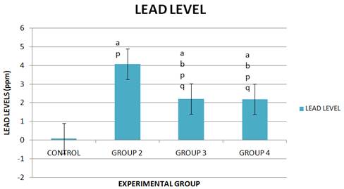
Figure 1. Lead levels in the experimental rats.
a- Significantly different from the Healthy Control Group (Group 1) at p<0.05
b- Significantly different from the Lead treated Group (Group 2) at p<0.05
p- Significantly different from the Healthy Control Group (Group 1) at p<0.01
q- Significantly different from the Lead treated Group (Group 2) at p<0.01
4. Discussion
The results of this study showed that normal saline has no effect on the body weight of the animals in the Control Group. The animals in the Lead acetate Control Group experienced progressive weight loss which was statistically significant in the fourth week. The weight loss might not be unconnected with the exposure of the animal to lead (Nwokocha et al, 2012). Lead is known to reduce protein synthesis possibly by damaging DNA and RNA (Shalan et al, 2005) and this might occur through base pair mutation, deletion and oxygen radical attack. Moreover, Lead disturbs intracellular calcium ion homeostasis (Simons, 1993) and damage the endoplasmic reticulum, which in turn results in reduction of protein synthesis. The animals in the group post treated with Basella alba L. showed a non significant weight gain when compared to the Healthy Control Group and those in the Pre treated Group also showed a non significant weight gain when compared to the Healthy Control Group after the first week This slight increase in weight could be due to the high calorie of protein contained in Basella alba (Duke and Ayensu, 1985).
Some biochemical parameters were measured in the serum of the experimental rats. Usually, when there is hepatotoxicity, enzymes like alkaline phosphatase, aspartate aminotransferase and alanine aminotransferase show a significant rise in their levels (Cornelius, 1979). In the rats treated with Lead Acetate alone there was a significant increase in their levels when compared to the rats in the Control Group (Group 1) only receiving normal saline, this is because there was damage to the liver tissues due to oxidative stress. There was a significant decrease in the level of these enzymes in the Lead exposed rats treated with Basella alba when compared with the Lead Control Group (Group 2), this might be due to the presence of antioxidants in Basella alba (Duke and Ayensu, 1985) and antioxidants protect against oxidative damage. Basella alba is known to contain the following antioxidants: basellasaponin A,B,C and D (Toshiyuki, 2001), flavonoid, betacyanin, phenolic acid and ascorbic acid (Duke and Ayensu, 1985; Daniel, 2006; Eliana et al, 2007) that are implicated to scanvage reactive oxygen species (ROS). The results of this study are in consonance with the results derived from a research done by Sharma et al, in 2010 where garlic was used as a source of antioxidants.
Glutathione has antioxidant effects in the body and during liver toxicity their levels in the liver and serum will decrease (Bechara, 2004). There was a significant decrease in glutathione in the rats treated with lead acetate when compared to the Control Group, this occurred due to the inhibition of the antioxidant activity of glutathione (Dickinson et al, 2003). The rats treated with Basella alba showed a significant increase in glutathione levels when compared to the Lead Acetate Control Group. This increase might due to the antioxidant capacity of basella alba (Duke and Ayensu, 1985; Kumar et al, 2013).
Also the amount of lead in the liver was determined and there was a higher concentration of lead seen in the Lead Acetate Control Group when compared with the Healthy Control Group. But, the Basella alba treated rats that were exposed to Lead Acetate had lower concentrations of lead when compared to the Lead Acetate Control Group. This indicates that Basella alba exhibited hepatoprotective activity against lead poisoning which is attributable to the basellasaponins contained in it (Toshiyuki, 2001). This study also supports the hepatoprotective potential of Basella alba earlier reported by Das et al, 2014 against paracetamol-induced hepatotoxicity in albino rats.
5. Conclusion
The results of this study show that basella alba is effective in reducing the accumulation of lead in liver tissues and can alleviate lead acetate induced changes in hepatic biochemical parameters. It has also shown that it is effective for hepatic protection and treatment of Lead poisoning and owing to the fact that it contains basellasaponins which is used as an antidote. Hence, consumption of Basella alba as a vegetable should be encourage in individuals especially in those who have been exposed to environmental toxicants like lead. It reduces toxic effects of lead in the body.
References
- Abalaka M., Oyewole O.A. and Kolawole A.R. (2011). Antibacterial Activities of Azadirachta Indica against Some Bacterial Pathogens. Journal of Microbiology Research; 1(2): 6-8.
- Abalaka M. E., and Oyewole O.A. (2011). Antibacterial Activities of Asmina triloba against Some Bacterial Pathogens. Journal of Microbiology Research; 1(1): 5-7.
- Aziz F.M., Maulood I.M. and Chawsheen M.A.H. (2012). Effects of melatonin, vitamin C and E alone or in combination on lead-induced injury in liver and kidney organs of rats. IOSR Journal of Pharmacy; 2(5): 13-18.
- Bamidele O, Akinnuga A.M., Olorunfemi J.O., Odetola O.A., Oparaji C.K. and Ezeigbo N. (2010). Effects of aqueous extract of Basella alba leaves on haematological and biochemical parameters in albino rats. African Journal of Biotechnology; 9(41):6952-6955.
- Bechara E.J.H. (2004). Lead poisoning and oxidative stress. Free Radical Biological Medicine; 36: 22.
- Budd P., Montgomery J., Cox A., Krause P., Barreiro B., Thomas R.G. (1998). The distribution of lead within ancient and modern human teeth: implications for long-term and historical exposure monitoring. Science Total Environment; 220(2-3):121-36.
- Cornelius C.E., (1979). Biochemical evaluation of hepatic function in dogs. Journal of American Animal Hospital Association; 15: 25-29.
- Cretacci Y. and Parsons P.J. (2010). Localized accumulation of lead within and among bones from lead-dosed goats. Environ Res; 110: 26-32.
- Daniel M. (2006). Medicinal plants: chemistry and properties. Science publishers, New Hampshire, USA. P. 198.
- Das S., Bandyopadhyay S., Ramasamy A., Mondal S. (2014). Evaluation of hepatoprotective activity of aqueous extract of leaves of Basella alba in albino rats. Natural Product Research; 29(11):1-6.
- DickinsonD.A., Moellering D.R., Iles K.E., Patel R.P., Levonen A.L. and Wigley A. (2003). Cytoprotection against oxidative stress and the regulation of glutathione synthesis. Biological Chemistry; 384: 527-537.
- Duke J.A. and Ayensu E.S. (1985). Medicinal Plants of China. Reference Publications, Inc. ISBN 0-917256-20-4.
- Eliana F.O., Paulo C.S., Milton C.C. (2007).Stability of anthocyamin in spainach vine (Basella rubra) fruits. Cien. Inv. Agr. 34(2): 115-120.
- Ellman G.C. (1959). Tissue sulfhydryl groups. Archives of biochemistry and biophysics; 82: 70-77.
- Flegal A.R. and Smith D.R. (1995). Measurements of environmental lead contamination and human exposure. Revolution of Environmental Contamination Toxicology; 143:1-45.
- Garaza A., Vega R. and Soto E. (2006).Cellular mechanisms of lead neurotoxicity. Medical Science Monitor; 12(3): 57-65.
- Grubben G.J.H. and Denton O.A. (2004). Plant Resources of tropical African vegetables. PROTA Foundation Wageningen; Backhuys Leiden (CTA) Wageningen; 4:103-110.
- Jachak S.M. and Saklani A. (2007). Challenges and opportunities in drug discovery from plants. Journal of Current Science; 92(1):1251-1257.
- Jackie T., Haleagrahara N. and Chakravarthi S. (2011). Antioxidant effects of Etlingera elatior flower extract against lead acetate -induced perturbations in free radical scavenging enzymes and lipid peroxidation in rats. BMC Research Notes; 4:67-75.
- Kayode A.A. and Kayode O.T. (2011). Some Medicinal Values of Telfairia occidentalis: A Review. American Journal of Biochemistry and Molecular Biology; 1:30-38.
- Khan M.S.H., Mostafa M.S., Hossain M.A. and Sayed M.A. (2008). Effect of garlic and vitamin B-complex in lead acetate induced toxicities in mice. Bangladesh Journal of Veterinary Medicine; 6(2): 203-210.
- Kumar S., Prasad A.K., Iyer S.V., Vaidya S.K. (2013). Systemic pharmacognostical, phytochemical and pharmacological review on an ethnomedicinal plant, Basella alba L. Journal of Pharmacognosy and Phytotherapy; 5(4):53-58.
- Newman D.J. (2003). Natural products as sources of new drugs over the period 1981-2002. Journal of Natural Products; 66: 1022-1037.
- Nriagu J.O. and Pacyna J.M. (1988). Quantitative assessment of worldwide contamination of air, water and soils by trace metals. Nature; 333:134-139.
- Nwokocha C.R., Nwokocha M.I., Owu D.U., Obi J., Olatunde B., Ebe C., Nwangwu O. and Iwuala M.O. (2012). Comparative analysis on the effect of palm oil (Elaeis guineensis) in reducing cadmium and lead accumulation in liver of Wistar rats. Pharmacognosy Research; 4(4): 214-218.
- Olaniyi T.A., Adeniran S.A., Adebayo L.A., Olusegun K.A. and Oyedeji T.A. (2013). Antioxidant and anti-lipid peroxidation potentials of the ethylacetate and chloroform extracts of basella alba leaves. Asian journal of natural and applied sciences; 2(2): 86.
- Reitman S. and Frankel A.S. (1957). A ccolorimetric method for the determination of serum glutamic oxaloacetic and glutamic pyruvic transaminase. American journal of clinical pathology; 28:53-56.
- Rodda R., Kota A., Sindhuri T., Kumar S.A. and Gnananath K. (2012). Investigation on anti-inflammatory property of Basella alba Linn leaf extract. International Journal of Pharmacy and Pharmaceutical Sciences; 4(1): 452-454.
- Shalan M.G., Mostafa M.S., Hassouna M.M., El-Nabi S.E. and El-Refaie A. (2005). Amelioration of lead toxicity on rat liver with vitamin C and silymarin supplements. Toxicology; 206: 1-15.
- Sharma A., Sharma V. and Kansal L. (2010). Amelioration of lead-induced hepatotoxicity by Allium sativum extracts in Swiss albino mice. Libyan journal of Medicine; 5: 4621.
- Sharma R.P. and Street J.C. (1980). Public health aspects of toxic heavy metals in animal feeds. Journal of the American Veterinary Medical Association; 177(2):149–153.
- Shi J., Arunasalam K., Yeung D., Kakuda Y., Mittal G. and Jiang Y. (2004). Saponins from edible legumes: chemistry, processing and health benefits. Journal of Med Food; 7(1): 67-78.
- Simons T. (1993). Lead-calcium interactions in cellular lead toxicity. Neurotoxicology; 14: 77-86.
- Toshiyuki M., Kazuhiro H. and Masayuki Y. (2001). Medicinal foodstuffs XXIII structures of new oleanane- type Triterpene- oligoglycosides, Basellasaponins A, B, C and D from the fresh aerial parts of Basella rubra L. Chemical and Pharmaceutical Bulletin; 49: 776 - 779.
- Woolf A.D. (2007). Update on the clinical management of childhood lead poisoning. Pediatric Clinics of North America; 54(2): 271–294.
- World Medical Association (2002). Americal Physiological Society.Guiding principles for research involving animals and human beings. Am. J. Physiol. Reg. Integr. Comp. Physiol., 283: 281-283.
- Xu Y., Li G., Han C., Sun L., Zhao R. and Cui S. (2005).Protective effect of Hippophae rhamnoides L. juice on lead-induced neurotoxicity in mice. Biological and Pharmacological Bulletin; 28:490-494.
- Yasmin H., Abul Kaisar M.D., Moklesur M.R.S., Mohammed S.R. and Mohammad A.R. (2009). Preliminary Antibacterial Activity of Some Indigenous Plants of Bangladesh, Dhaka University. Journal of Pharm Science; 8(1): 61-65.


