International Journal of Plant Science and Ecology, Vol. 1, No. 5, October 2015 Publish Date: Jul. 23, 2015 Pages: 190-195
Antioxidant and Hepatoprotective Potential of Ethanolic Leaves Extract of Jatropha gossypifolia
Sachin Jain1, *, G. P. Choudhary2, D. K. Jain1
1College of Pharmacy, IPS Academy, Indore, India
2School of Pharmacy, Devi Ahilya Vishwavidyalaya, Indore, India
Abstract
Jatropha gossypifolia Linn. (JG) has been traditionally used in the Ayurvedic system of medicine as a chief ingredient of many polyherbal formulations as antioxidant. To evaluate the antioxidant and hepatoprotective activity of ethanolic extract of Jatropha gossypifolia leaves. Animals were pre-treated with ethanolic plant extract for 7 days and then toxicity was induced with a single CCl4 intraperitoneal injection. Pre-treatment with 500 mg/kg (p.o.) of ethanolic extract of Jatropha gossypifolia improved the glutathione (oxidized), SOD, catalase, and peroxidase lipid peroxidation levels significantly as compared to control group. Liv 52 was used as standard reference and exhibited significant hepatoprotective activity against carbon tetrachloride induced hepatotoxicity in rats. The biochemical observations were supplemented with histopathological examination of rat liver sections. The results of this study strongly indicate that Jatropha gossypifolia leaves have potent hepatoprotective action against carbon tetrachloride induced hepatic damage in rats. Ethanolic extract was found more potent hepatoprotective. Meanwhile, in vivo antioxidant activities were also screened which were positive for ethanolic extracts. On the basis of this study we conclude that ethanolic extract of Jatropha gossypifolia possesses in vivo antioxidant activity and can be employed in protecting tissue from oxidative damage and stress.
Keywords
Jatropha gossypifolia, Liv 52, CCl4, Antioxidant Activity, Superoxide Dismutase, Hepatoprotective
Received: May 26, 2015
Accepted: June 20, 2015
Published online: July 23, 2015
@ 2015 The Authors. Published by American Institute of Science. This Open Access article is under the CC BY-NC license. http://creativecommons.org/licenses/by-nc/4.0/
Contents
1. Introduction 2. Materials and Methods 2.1. Collection of Plant 2.2. Chemicals and Drugs 2.3. Preparation of Extracts 2.4. Preliminary Phytochemical Screening 2.5. Animals 2.6. Acute Toxicity Test 2.7. CCL4 Induced Hepatotoxicity 2.8. Biochemical Estimation 2.9. Histopathological Studies 2.10. Statistical Analysis 3. Result 3.1. Phytochemicals Investigation 3.2. Acute Toxicity Studies 3.3. In-Vivo Antioxidant Activity 3.4. Histopathological Observations 4. Discussion 5. Conclusion
1. Introduction
Jatropha gossypifolia (Euphorbeacea) a common garden plant in tropical countries has been used as a traditional medicine. Plants are well known as a major source of modern medicines. From ancient times, humans have utilized plants for the treatment or prevention of diseases, leading to the dawn of traditional medicine. Jatropha gossypifolia is one of the genera that are used in Chinese, Ayurvedic and Thai traditional medicine for the treatment of fever, pain and dysentery [1].
Literature reveals that, the carbonyl groups are responsible for free radical scavenging activity. Free radicals are atoms or groups of atoms with an odd number of electrons and can be formed when oxygen interacts with certain molecules. To prevent free radical damage, the body has a defense system of antioxidants [2,3]. Antioxidants are able to give free radicals, which becomes a companion to their unpaired electron, thus eliminating the threat of gene alteration leading to cancer [2,3]. Medicinal plants have attracted attention of not only professionals from various systems of medicines, but also the scientific community belonging to different disciplines [4,5]. In recent years, these have been a great interest in herbal remedies for the treatment of number of ailments. In continuation of search in potential free radical, scavenging agents [6] the present investigation was aimed to determine antioxidant activity of Jatropha gossypifolia leave (Linn.) properties to strengthening the immune system of the body which helps to overcome cancer.
The antioxidant activity of Du-Zhong (Eucomnia ulmoides) [7], ear mushrooms and anise (Pimpenella anisum L.) seed were found to correlate with the phenolic compounds. Studies on local plants such as turmeric (Curcuma domestica), betel leaf (Piper betel), pandan leaf (Panadanus odorus), asam gelugur (Garnicia atroviridis), mengkudu (Morinda citrifolia), pegaga (Centella asiatica), ginger (Zingiber officinale), cassava shoot (Manihot asculenta), kesum (Polygonum minus) and selom (Oenathe javanica) also exhibit good antioxidant activity. The antioxidative properties of some vegetables and fruits are partly due to the low molecular weight phenolic compounds which are known to be potent as antioxidants [8].
2. Materials and Methods
2.1. Collection of Plant
Leaves of Jatropha gossypifolia (Linn.) were collected from railway track and road side places of Indore MP and voucher specimen (COPIPS/T/098/2010) has been deposited. The authentication was done by Prof. S. R. Upadhyaya Professor, Govt. Post graduate College, Indore (M.P.) India.
2.2. Chemicals and Drugs
Carbon tetra chloride, Potassium chloride, Ellman’s reagent, hydroxylamine.
2.3. Preparation of Extracts
The leaves of Jatropha gossypifolia were collected and shade dried. The dried leaves were coarse powdered and the powder was packed in to Soxhlet apparatus and extracted with ethanol (64.5 – 65.5°C). The extract was concentrated under reduced pressure (bath temp 50°C). The dried extracts were stored in airtight container.
2.4. Preliminary Phytochemical Screening
Tests for common phytochemicals were carried out by standard methods [9,10].
2.5. Animals
Albino rats of Wister strain, weighing 100–150 g, kept on normal diet (Ashirwad Industries, Punjab) and water ad libitum, were divided into six groups of six animals each. Before starting the experiment, permission from the Institutional Animal Ethics Committee was obtained.
2.6. Acute Toxicity Test
The acute toxicity study for ethanolic and aqueous extracts (decocted and macerated) of J. gossypifolia leaves were performed using albino mice and rats. The animals were fasted overnight prior to the experiment and maintained under standard conditions. All the extracts were administrated orally in increasing dose and found safe up to dose of 2000 mg/kg for all extracts.
2.7. CCL4 Induced Hepatotoxicity
Group-I animals served as normal control, treated with vehicle (gum acacia 3% solution). Group-II animals served as toxic control, treated with CCl4 in a single dose of 1.5 ml/kg, i.p., to produce acute toxicity. Group III served as a standard group, and was administered Liv-52 (Anti-peroxidative and Hepatoprotective) in a dose of 56 mg/kg, p.o. Group-IV, V and -VI animals were treated with daily doses of 100, 300 and 500 mg/kg, p.o., respectively, of ethanolic extract of J. gossypifolia for 7 days. The animals of Groups III–VI were given single dose of CCl4, 1.5 ml/kg, i.p., 6 h after the last treatment. On day 8 the rats were sacrificed by carotid bleeding and liver was rapidly excised, rinsed in ice-cold saline, and a 10% w/v homogenate was prepared using 0.15M KCI, centrifuged at 800 g for 10 min at 4°C. The supernatant obtained was used for the estimation of catalase, peroxidase, and other enzymes. Further, the homogenate was centrifuged at 1000 rpm for 20 min at 4°C and the supernatant was used for biochemical estimation.
2.8. Biochemical Estimation
2.8.1. Estimation of Glutathione
Glutathione was estimated using Ellman’s reagent (5, 5- dithiobis-(2-nitrobenzoic acid) [DTNB]). The sulphydryl groups present in glutathione forms a colored complex with DTNB, which was measured by colorimeter at 412 nm. The amount of glutathione was determined using its molar extinction coefficient of 13600/m/cm and expressed in terms of μmol/mg of protein [11].
2.8.2. Estimation of SOD
Estimation of SOD was done by auto oxidation of hydroxylamine at pH 10.2, which was accompanied by reduction of NBT, and the nitrite produced in the presence of EDTA was detected colorimetrically. One enzymatic unit of SOD is the amount in the form of proteins present, 10% liver homogenate required to inhibit the reduction of 24 mM NBT by 50% and is expressed as units per milligram of protein.
2.8.3. Estimation of Catalase
Catalase activity was estimated by determining the decomposition of H2O2 at 240 nm in an assay mixture containing phosphate buffer. One international unit of catalase utilized is that amount that catalyzes the decomposition of 1 mM H2O2/min/mg of protein at 37°C. Catalase activity was calculated using the millimolar extinction coefficient of 0.07 and expressed in terms of micromole per minute per milligram of protein.
2.8.4. Estimation of Peroxidase
Peroxidase estimation is based on per-iodide formation. Per-iodide can be spectrophotometrically determined at 353 nm, and this is directly proportional to the peroxidase concentration in the reaction mixture containing approximate amounts of H2O2 and enzyme [12]. One unit of peroxidase activity is defined as the change in absorbance per minute and expressed in terms of units per milligram of protein.
2.8.5. Assay of Lipid Peroxidation Using Mouse Brain Homogenates
The brains of young adult male mice were dissected and homogenized with a Polytron in ice-cold Tris- HCl buffer (20 mM, pH 7.4) to produce a 1/10 homogenate. The homogenate was centrifuged at 12000g for 15 min at 4 °C, and the supernatant was used for in vitro lipid peroxidation assay. A 1 mL aliquot of the supernatant was incubated with the test samples (final extract concentrations in the assays were 1, 5, 10, and 50 µg/mL, respectively) in the presence of 10 µM FeSO4 and 0.1 mM ascorbic acid at 37 °C for 1 h. The reaction was terminated by the addition of 1.0 mL of trichloroacetic acid (TCA; 28%, w/v) and 1.5 mL of thiobarbituric acid (TBA 1%, w/v), followed by heating at 100 °C for 15 min. After centrifugation to remove precipitated protein, the absorbance of the malondialdehyde (MDA)-TBA complex was measured at 532 nm. (+)-Catechin was used as a positive control. The inhibition ratio (percent) was calculated from the following equation:
![]()
2.9. Histopathological Studies
The liver tissue was dissected out and fixed in 10% formalin, dehydrated in gradual ethanol (50–100%), cleared in xylene, and embedded in paraffin. Sections were prepared and then stained with hematoxylin and eosin (H–E) dye for photomicroscopic observation, including cell necrosis, fatty change, hyaline regeneration, ballooning degeneration.
2.10. Statistical Analysis
All analyses were run in triplicates. Data were analyzed by an analysis of variance (ANOVA). Statistical analysis was performed by one-way ANOVA followed by post hoc Dennett’s test.
3. Result
3.1. Phytochemicals Investigation
It was found that ethanolic extract contained carbohydrates, amino acids, steroids, flavonoid, alkaloids, glycosides and tannins.
3.2. Acute Toxicity Studies
Ethanolic and aqueous extracts did not show any sign and symptoms of toxicity and mortality up to 2000 mg/kg dose.
3.3. In-Vivo Antioxidant Activity
Phytochemical screening of the plant shows the presence of flavanoids in ethanolic extract. Therefore, the present study was undertaken to assess the in vivo antioxidant effects of ethanolic extract of J. gossypifolia on CCl4 induced hepatotoxicity in rats. The result showed that the activities of glutathione, SOD, catalase and peroxidase in group treated with CCl4 declined significantly than that of normal group. Co-administration of ethanolic extract of J. gossypifolia at a dose of 100, 250 and 500 mg/kg for 7 days markedly prevented these CCl4 induced alteration and maintained enzymes level near to normal values. Standard (Liv 52) treated group also significantly increased the level of glutathione, SOD, Catalase and peroxidase in CCl4 induced hepatotoxic rats. Lipid peroxidation (LPO) levels of liver homogenate were summarized in Table 1.
Lipid peroxidation decreased towards the normal value in ethanolic extract treated carbon tetrachloride intoxicated rats. The antioxidant activity or the inhibition of the generation of free radical is important in the protection against CCl4-induced liver lesion. The level of lipid peroxide is a measure of membrane damage alterations in its structure function. The level of MDA, which is one of the end products of lipid peroxidation in liver tissue, was found to be high in CCl4 control group (shown in Table 1) implying enhanced lipid peroxidation leading to tissue damage failure of antioxidant defense mechanisms against free radicals.
3.4. Histopathological Observations
Histology of the liver sections of control animals (Group I) showed normal hepatic cells with well-preserved cytoplasm, prominent nucleus, nucleolus and visible central veins. The liver sections of CCl4-intoxicated rats showed massive fatty changes, necrosis, ballooning degeneration and broad infiltration of the lymphocytes and the loss of cellular boundaries. The histological architecture of liver sections of the rats treated with ethanolic extracts showed more or less normal lobular pattern with a mild degree of fatty change, necrosis and lymphocyte infiltration almost comparable to the control and Liv 52 treated groups (Fig. 1).
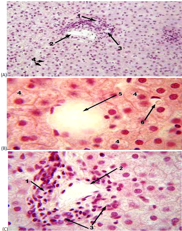
Fig. 1. (A) Liver sections of normal control rats showing: normal hepatic cells with well-preserved cytoplasm; well brought out central vein; prominent nucleus and nucleolus. (B) Liver section of CCl4 treated rats showing: massive fatty changes, necrosis, ballooning degeneration, and broad infiltration of the lymphocytes and the loss of cellular boundaries. (C) Liver section of rats treated with CCl4 and extracts showing: well brought out central vein, hepatic cell with well-preserved cytoplasm, prominent nucleus and nucleolus (H, heptocytes; N, nucleus; CV, central vein; CP, cytoplasm; B, ballooning; NC, necrosis).
Table 1. Effect of ethanolic extract of Jatropha gossypifolia on biochemical parameters.
| Treatment | Glutathione SOD Catalase Peroxidase (μmol/mg of protein) | SOD (u/mg of protein) | Catalase (uM/min/mg of protein) | Peroxidase (u/mg of protein) | Lipid Peroxidation [MDA content (in nano mole/milligram)] |
| Normal control(Vehicle treated) | 18.5 ± 0.094 ** | 32±0.02** | 11±0.03** | 84±0.09** | 20.78 ± 0.757 |
| Hepatotoxic Control (CCl4 Treated) | 8.4 ± 0.7 | 19±0.01 | 1.1±0.01 | 13±0.02 | 99.31±5.59** |
| Standard Liv 52 | 16.3 ± 0.07** | 31±0.01** | 8.5±0.1** | 67±0.15** | 39.40±4.0398* |
| EEJG 100 mg/kg | 9.9 ± 0.06 | 23±0.02 | 1.6±0.2 | 15±0.06 | 15.39 ± 0.479* |
| EEJG 250 mg/kg | 12.9 ± 0.1* | 25±0.01 | 1.8±0.4 | 17±0.03 | 18.34 ± 0.348 |
| EEJG 500 mg/kg | 15±0.9** | 27±0.05* | 5.1±0.4** | 63±0.06** | 75.28±7.35* |
EEJG: Ethanolic Extract of Jatropha gossypifolia
Values are in Mean±SEM.
Where* p<0.05 and **p<0.01
4. Discussion
The traditional medicine all over the world is nowadays revalued by an extensive activity of research on different plant species and their therapeutic principles. Experimental evidence suggests that free radicals (FR) and reactive oxygen species (ROS) can be involved in a high number of diseases [13]. As plants produce a lot of antioxidants to control the oxidative stress caused by sunbeams and oxygen, they can represent a source of new compounds with antioxidant activity.
In the present study, the ethanolic extract was selected as they contain alkaloids, glycosides, saponins, tannins, flavonoids and phenolic compounds. This may have active constituents for producing the free radical scavenging effect.
Free radicals are produced under certain environmental condition and during normal cellular function in the body. These molecules are missing an electron, giving them an electric charge. To neutralize this charge, free radicals try to withdraw an electron from, or donate an electron to, a neighboring molecule. Other antioxidants works against the molecules that form free radicals, destroying them before they can begin the domino effect that leads to oxidative damage [14].For example, certain enzymes in the body, such as superoxide dimutase, work with other chemical to transfer free radical into harmless molecules. Vitamin C an antioxidant that may prevent cataracts and cancers of the stomach throat, mouth, and pancreas [15]. It may also prevent the oxidation of LDL cholesterol, lowering the risk of heart disease. This investigation revealed that the Jatropha gossypifolia contains pharmacologically active substance (s) such as alkaloids, glycosides, saponins, tannins, flavonoids and phenolic compounds, which are responsible for the antioxidant activity.
5. Conclusion
As ethanolic extract of J. gossypifolia in the dose of 500 mg/kg, p.o., has improved the glutathione, SOD, catalase, and peroxidase levels significantly, which were comparable with Liv 52. On the basis of the study we conclude that ethanolic extract of J. gossypifolia possesses in-vivo antioxidant activity and can be employed in protecting tissue from oxidative stress.
References
- Geronikaki, A., Litina, D. H., Chatziopoulos C, Soloupis, G. (2003). Synthesis and Biological Evaluation of New 4, 5-Disubstituted-Thiazolyl Amides, Derivatives of 4-hydroxy- piperidine or of 4-N-Methyl piperazine. Molecules 8: 472-479.
- Wasana, P., Anchalee, P., Nipon, C., Siriporn, C. (2008). Ethnobotany & ethnopharmacology of Tabernaemontana Divaricata, Indian J Med Res 127: 317- 335.
- Patil, S., Jolly, C. I., Narayanan, S. (2003). Free Radical Scavenging Activity of Acaci catechu and Rotula aquitica implication in Cancer Therapy, Indian Drugs 40: (6), 328-332.
- Sharma, P. C., Yelane, M. P., Dennis, T. J. (2002). Data base on Medical Plants used in Ayurveda. New Delhi: Central council for research in Ayurveda and Siddha 1: 45-47.
- Chhajed, M. R., Khedekar, P. B., Mundhey, A. S. (2007). Synthesis and free radical scavenging activity of some 1, 3, 4-thiazole derivative. Indian journal of heterocyclic chemistry 16: 259-262.
- Khandelwal, K. R. (2000). Practical Pharmacognosy techniques and experiments. (2nd ed). Nirali Prakashan, Pune, 34-38.
- Kokate, C. K. (1999). Practical Pharmacognosy. (4th ed). vallabh Prakashan, New Delhi, 230-34.
- Siddique, N. A., Mujeeb, M., Najmi, A. K., Akram, M., (2010). Evaluation of antioxidant activity, quantitative estimation of phenols and flavonoids in different parts of Aegle marmelos. African Journal of Plant Science 4: 001–005.
- Kuriakose, G. C., Kurup, M. G., (2011). Antioxidant and antihepatotoxic effect of Spirulina laxissima against carbon tetrachloride induced hepatotoxicity in rats. Food Function 2: 190–196.
- Bumrela, S. B., Naik, S. R., (2011). Identification of β-carotene and β-sitosterol in methanolic extract of Dipteracanthus patulus (Jacq) nees and their role in antimicrobial and antioxidant activity. International Journal of Phytomedicine 3: 204–215.
- Bagul, M. S., Niranjan, S. K., Rajani, M. (2005).Evaluation of free radical scavenging properties of two classical polyherbal formulations, Indian Journal of Experimental Biology 43:732-36.
- Marzouk, A. M., (2009). Hepatoprotective triterpenes from hairy root cultures of Ocimum basilicum L. Zeitschrift fur NaturforschungTeilC: BiochemieBiophy- sik BiologieVirologie 64: 201–209.
- Usha, S. S., Vilasrao, J. K., Rumi, G., (2008). Hepatoprotective activity of livo bond, a polyherbal formulation against carbon tetrachloride induced hepatotoxicity in rats. International Journal of Pharmaceutics 4: 472–476.
- D’Mello, P. M., Jadhav, M. A., Jolly, C. I. (2000). Free Radical Scavenging Activity of Syzygium Cumini and ficus bengalensis Plants Used in Ayurveda for Diabetes Mellitus. Indian Drugs 37: (11), 518-520.
- Atawodi, S. E., Onaolapo, G. S., (2010). Comparative invitro antioxidant potential of different parts of Ipomoea asarifolia, Roemer and Schultes, Guiera senegalensis, J. F. Gmeland Anisopus mannii N. E. Brown Brazilian Journal of Pharmaceutical Sciences 46: 245–250.


