International Journal of Plant Science and Ecology, Vol. 1, No. 3, June 2015 Publish Date: May 18, 2015 Pages: 113-118
Evaluation of Antibacterial Potential of Stem Bark of Ethnomedicinal Tree Pittosporum dasycaulon Miq
Bince Mani, T. Dennis Thomas*
Postgraduate and Research Department of Botany, St. Thomas College, Palai, Arunapuram (P.O.), Kottayam, Kerala, India
Abstract
The ethnic people indigenous to Southern Western Ghats using stem bark of Pittosporum dasycaulon Miq. in their medicinal preparations to treat various skin diseases, leprosyand bronchitis and also used as antifungal and antibacterial agent. The dried stem bark of P. dasycaulon was serially extracted with petroleum ether, chloroform, acetone, methanol and water. The extracts were concentrated and their antibacterial activity was detected against thirteen bacterial strains by disc diffusion method. Minimum inhibitory concentration (MIC) of each extract was also determined. Chloroform and acetone extracts displayed a potential antibacterial activity, against all the tested gram-positive bacteria and eight gram-negative bacteria, compared to petroleum ether, methanol and aqueous extracts. Among the different fractions chloroform extract was most effective with maximum zone of inhibition (23.33±0.58 mm) and lowest MIC value (≤0.325 mg/ml) against Enterobacter aerogens. The aqueous extract was least effective against all the tested bacteria. The results of this investigation suggest that stem bark of Pittosporum dasycaulon can be used to discover broad spectrum antibacterial principles for developing new pharmaceuticals to control studied and many other human pathogenic bacteria responsible for severe illness.
Keywords
Pittosporum dasycaulon, Stem Bark, Ayurveda, Disc Diffusion, Minimum Inhibitory Concentration
Received: April 8, 2015 / Accepted: April 26, 2015 / Published online: May 15, 2015
@ 2015 The Authors. Published by American Institute of Science. This Open Access article is under the CC BY-NC license. http://creativecommons.org/licenses/by-nc/4.0/
1. Introduction
The frequency of life-threatening infections caused by pathogenic microorganisms has increased worldwide and is becoming an important cause of morbidity and mortality in immunocompromised patients in developing countries1. The increasing prevalence of multi-drug resistant strains of bacteria and the recent appearance of strains with reduced susceptibility to antibiotics raised the spectre of ‘untreatable’ bacterial infections and adds urgency to the search for new infection-fighting agents2-3. Over the past twenty years, there has been a lot of interest in the investigation of natural materials as sources of new antibacterial agents4-5. Plant based antimicrobials represent a vast untapped source for medicines and further exploration of plant antimicrobials needs to occur. Antimicrobials of plant origin have enormous therapeutic potential6. Different extracts from traditional medicinal plants have been tested and many reports have show the effectiveness of traditional herbs against microorganisms, as a result, plants are one of the bedrocks for modern medicine to attain new principles7-9.
Primitive people have used plant derived preparations to cure a variety of human ailments including skin diseases10. In India, the use of herbal drugs is an important component of the traditional system of medicine. Many Indian plants have been used from time immemorial to treat various diseases and infections in traditional medicinal systems of India such as Siddha, Ayurvedha, Naturopathy, etc11-12. Even today, 85% of Indians use higher plants as effective antimicrobials for the treatment of various diseases13. A vast number of medicinal plants which are used in Indian traditional system have not been studied for their antibacterial properties scientifically including species of Pittosporum. In India, the bark of Pittosporum genus is used as anti-inflammatory, antispasmodic, antidote and for narcotic effects. Its bark is found to be useful in chronic bronchitis, bacterial and fungal diseases and is also administered in leprous infections and rheumatic swellings14-16.
Pittosporum dasycaulon Miq. is a small evergreen tree, belongs to the family Pittosporaceae. The plant is distributed in the moist deciduous and evergreen forests of southern and central parts of Western Ghats above 800 m from mean sea level (MSL)16. Bark is bitter, aromatic and has powerful antioxidant potential17. The bark and stem of P. dasycaulon have been used in folk medicine as an antibacterial and antifungal agent to treat infection14. Essential oil obtained from the stem bark of this plant shows antibacterial activity18.
As an important wild source of medicine Pittosporum dasycaulon Miq. is yet to receive sufficient attention. Furthermore, little information about the antibacterial effect of thisspecies is available. Therefore, the present investigation was aimed at evaluating the antibacterial potential of the extracts of stem bark of Pittosporum dasycaulon Miq. an ethnomedicinal plant endemic to Western Ghats16.
2. Material and Methods
2.1. Plant Material and Preparation of Extracts
Stem bark of Pittosporum dasycaulon Miq. was collected from Southern Western Ghats, Kerala, India, in 2010. The species was identified by Dr. S. John Britto, The Rapinat Herbarium and Centre for Molecular Systematics and voucher specimen (RHT58608) has been deposited in The Rapinat Herbarium, St. Joseph’s College, Tiruchirappalli, Tamil Nadu, India.
The plant material brought to the laboratory and washed with distilled water to remove the adhering dust particles. Extracts were prepared from shade dried (29 ± 2°C) and ground sample of the plant part collected. About100 g powdered plant material was subjected to successive extraction using Soxhlet apparatus with petroleum ether, chloroform, acetone, methanol and water. After extraction with a solvent, the powdered plant material was air dried overnight at room temperature for complete evaporation of extracting solvent before subsequent extraction with other solvent. The individual extracts were collected, filtered and concentrated using rotary evaporator (Buchi) under reduced pressure and dried in a vacuum oven which was collected in sterile container and stored at 4°C in air tight containers for further studies. Stock solutions of crude extracts were prepared by dissolving the dried extracts in dimethyl sulphoxide (DMSO) to obtain a final concentration of 200 mg/ml.
2.2. Test Microorganisms
The bacterial strains used in the study were three Gram-positive, namely, Streptococcus haemolyticus (MTCC442), Bacillus cereus (MTCC430) and Staphylococcus aureus (MTCC87), and ten Gram-negative, namely, Vibrio parahaemolyticus (MTCC451), Vibrio cholera (MTCC3904), Salmonella paratyphi (MTCC735), Enterobacter aerogens (MTCC111), Salmonella typhi (MTCC733), Escherichia coli (MTCC433), Proteus vulgaris (MTCC426), Klebsiella pneumoniae (MTCC3384), Pseudomonas aeruginosa (MTCC741) and Serratia marcescens (MTCC86). All the tested strains are reference strains, and were collected from Microbial Type Culture Collection and Gene Bank, Institute of Microbial Technology, Sector 39-A, Chandigarh-160036, India.
2.3. Preparation of Inoculums
Suspension of organism was prepared as per McFarland nephelometerstandard19. A 24 hour old culture was used for the preparation of bacterial suspension. Suspension of organism was made in a sterile isotonic solution of sodium chloride (0.9% w/v) and the turbidity was adjusted such that it contained approximately 1.5*108 colony forming units/ml (CFU/ml). It was obtained by adjusting the optical density of the bacterial suspension to that of a solution of 0.05 ml of 1.175% w/v of barium chloride and 9.95 ml of 1% v/v (0.18 mol/L) sulphuric acid.
2.4. Antibacterial Assay
The antibacterial activity of different extracts was carried out by disc diffusion method20, recommended by NCCLS, using 25 μl of standardized suspension of test bacteria (1.5*108CFU/ml) spread on Mueller-Hinton agar (MHA, pH 7.3 ± 0.1) plates. The discs (6 mm in diameter) were impregnated with 10 μl (2 mg/disc) of the test compound, followed by air-drying and were placed on seeded agar plates. Negative control was prepared using the same solvent (DMSO) to dissolve the plant extracts. Amoxicillin (30 μg/disc) was used as positive control to determine the sensitivity of bacterial strain. The plates were incubated at 37°C for 24 h. Antimicrobial activity was evaluated by measuring the zones of inhibition against the tested bacteria. Each assay was carried out in triplicate.
2.5. Determination of Minimum Inhibitory Concentration (MIC)
The dilution test was performed to determine minimum inhibitory concentrations (MICs) using the standard procedure as described by Jorgensen et al21., with slight modifications. One hundred microliters of Mueller-Hinton broth (MHB) were added in each well of a microtiter plate. The stock solutions of each extracts were prepared in DMSO (200 mg/ml) and 100 μl aliquot of stock solution of crude extract was added in to the third well, and subsequently two-fold serially diluted up to eleventh well with MHB. The inoculums suspension (20 μl) of bacterial strain was then added in each well containing crude extract and MHB except second well, which was treated as sterility control. The final concentrations of the extract were 83.3, 41.7, 20.8, 10.4, 5.2, 2.6, 1.3, 0.65 and 0.325 mg/ml. The negative (first well) and positive (last well) controls were also performed using DMSO and amoxicillin respectively. Duplicate wells were run for each concentration of P. dasycaulon stem bark extracts. The plates were incubated at 37°C for 24 h, and the turbidity was measured at 620 nm using the microplate reader (iEMS Reader MF, Labsystems). The lowest concentration that inhibited visible growth of the tested organisms was recorded as the MIC.
3. Results
The antibacterial activities of P. dasycaulon stem bark extracts were examined against thirteen clinical pathogens causing severe respiratory and skin illness in human. The results of the disc diffusion test indicated that crude extracts of P. dasycaulon stem bark showed different degrees of growth inhibition, depending on the bacterial strains (Figure 1). As per the investigation, four extracts exhibited antibacterial activity. The chloroform extract showed the highest antibacterial activity followed by acetone, petroleum ether and methanol extracts. Among the thirteen bacterial strains were tested, nine (69.2%) showed susceptibility towards petroleum ether extract. The chloroform and acetone extracts inhibited the growth of eleven (84.6%) bacterial strains tested, while methanol extract inhibited the growth of four bacterial strains tested (31%). Aqueous extract did not show any inhibition of all the bacteria tested (Table 1).
Table 1. Diameter of zones of inhibition (mm) stem bark extracts of Pittosporum dasycaulon against test bacteria.
| Bacteria | a | b | c | d | e | Amoxicillin |
| Vibrio parahaemolyticus (-) | 12.00±1.00 | 18.33±0.58 | 14.33±0.58 | 13.33±0.58 | * | 27.66±0.57 |
| Pseudomonas aeruginosa (-) | 16.33±1.52 | 20.33±0.58 | 17.70±0.58 | * | * | 33.00±0.00 |
| Bacillus cereus (+) | 10.33±0.57 | 14.33±1.52 | 11.66±1.53 | * | * | 35.66±1.15 |
| Enterobacter aerogens(-) | 19.00±1.00 | 23.33±0.58 | 19.66±0.58 | 11.33±1.52 | * | 25.66±0.57 |
| Salmonella paratyphi (-) | 13.33±1.15 | 15.00±1.73 | 15.00±1.00 | 09.00±1.73 | * | 40.33±0.57 |
| Vibrio cholerae (-) | * | 12.66±2.08 | 10.33±1.52 | * | * | 30.00±1.00 |
| Staphylococcus aureus (+) | 08.33±0.57 | 14.00±2.00 | 14.00±1.00 | * | * | 34.33±1.15 |
| Serratiamarcescens (-) | 11.66±2.08 | 13.66±1.53 | 14.66±1.15 | * | * | 41.0±0.00 |
| Streptococcus haemolyticus (+) | * | 17.00±1.00 | 11.33±1.53 | * | * | 36.66±0.57 |
| Proteus vulgaris (-) | 17.33±0.57 | 21.33±0.58 | 16.33±0.58 | 08.66±1.15 | * | 24.33±0.57 |
| Klebsiellapneumoniae (-) | 08.66±0.57 | 14.66±0.57 | 11.33±1.52 | * | * | 32.33±1.15 |
| Escherichia coli (-) | * | * | * | * | * | 30.33±0.57 |
| Salmonella typhi (-) | * | * | * | * | * | 36.00±1.00 |
Note: Values are presented as mean± standard error of triplicate experiments.
a petroleumether extract, b chloroform extract, c acetone extract, d methanol extract, e aqueous extract; (+) gram positive, (-) gram negative; * no zone of inhibition observed.
Chloroform extract showed the broadest antibacterial activity by inhibiting the growth of Streptococcus haemolyticus, Bacillus cereus, Staphylococcus aureus (gram-positive), Vibrio parahaemolyticus, Pseudomonas aeruginosa, Enterobacter aerogens, Salmonella paratyphi, Vibrio cholerae, Serratia marcescens, Proteus vulgaris, and Klebsiella pneumonia (gram-negative) and the diameter of zones of inhibition were 17.0±1.0, 14.33±1.52, 14.0±2.0, 18.33±0.58, 20.33±0.58, 23.33±0.58, 15.0±1.73, 12.66±2.08, 13.66±1.53, 21.33±0.58 and 14.66±0.57 mm respectively. Acetone extract showed potent inhibitory effect against all the tested bacteria except Escherichia coli and Salmonella typhi (Table 1). Petroleum ether extract exhibited inhibitory effect against Bacillus cereus, Staphylococcus aureus, Vibrio parahaemolyticus, Pseudomonas aeruginosa, Enterobacter aerogens, Salmonella paratyphi, Serratia marcescens, Proteus vulgaris, and Klebsiella pneumonia and the diameter of zones of inhibition were depicted in table 1. V. parahaemolyticus, E. aerogens, S. paratyphi and P. vulgaris were susceptible for methanol extract. V. parahaemolyticus, P. aeruginosa, E. aerogens and P. vulgaris were found to be more susceptible as compared to other bacteria. In all cases, the activity of the extract was compared with standard antibiotic amoxicillin (Figure 1). The results revealed that among the thirteen pathogenic bacteria Salmonella typhi and Escherichia coli were insensitive to extracts of stem bark of P. dasycaulon.
Table 2 displays the minimum inhibitory concentrations (MIC) of different extracts against the tested organisms. The MIC values of four extracts were ranged from ≤0.325 to 83.3 mg/ml. Petroleum ether extract exhibited MIC ranging from ≤0.325 to 41.7 mg/ml against the tested organisms, except E. coli and S. typhi. The MIC analysis showed that the chloroform extract was highly active in comparison to other extracts, which inhibited the series of study organisms at a low concentration range (≤0.325 – 2.6 mg/ml) except E. coli and S. typhi (41.7 mg/ml). Acetone extract inhibited the growth of all bacteria tested at concentrations ranging from ≤0.325 to 83.3 mg/ml. The growth of S. haemolyticus, B. cereus, S. aureus, V. parahaemolyticus, V. cholerae, E. aerogens, P. vulgaris, P. aeruginosa and S. marcescens were inhibited by methanol extract at higher concentrations and MIC values were 5.2 to 83.3 mg/ml, whereas none of the bacteria were inhibited by the aqueous extract of stem bark of P. dasycaulon at any concentrations tested.
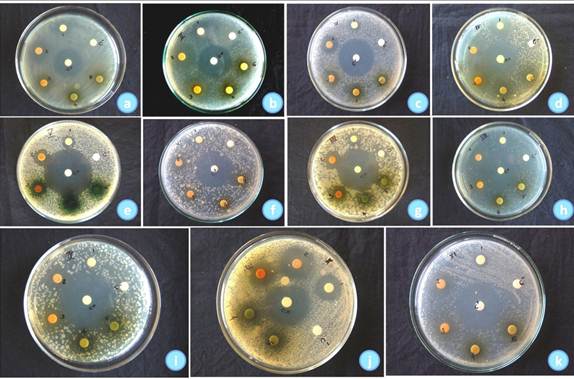
Figure 1. Antibacterial activity of various extracts of Pittosporum dasycaulon Miq. stem bark against Vibrio parahaemolyticus.
(a), Pseudomonas aeruginosa (b), Bacillus cereus (c), Enterobacter aerogens (d), Salmonella paratyphi (e), Vibrio cholerae (f), Staphylococcus aureus (g),
Serratia marcescens (h), Streptococcus haemolyticus (i), Proteus vulgaris (j) and Klebsiella pneumoniae (k). 1. Petroleum ether extract, 2. chloroform extract,
3. acetone extract, 4. methanol extract, 5. aqueous extract, C+: positive control (Amoxicillin) and C - : negative control (Dimethyl sulphoxide).
Table 2. Minimum inhibitory concentration (MIC) of different solvent extracts of Pittosporum dasycaulon stem bark against various microorganisms.
| Bacteria | a | b | c | d | e |
| Vibrio parahaemolyticus (-) | 0.65 | ≤0.325 | 0.65 | 5.20 | * |
| Pseudomonas aeruginosa (-) | 0.325 | ≤0.325 | 0.325 | 10.40 | * |
| Bacillus cereus (+) | 5.20 | 2.60 | 2.60 | 20.80 | * |
| Enterobacter aerogens(-) | ≤0.325 | ≤0.325 | ≤0.325 | 5.20 | * |
| Salmonella paratyphi (-) | 2.60 | 0.65 | 0.65 | 20.80 | * |
| Vibrio cholerae (-) | 41.70 | 0.65 | 5.20 | 83.30 | * |
| Staphylococcus aureus (+) | 10.40 | 0.65 | 0.65 | 20.80 | * |
| Serratia marcescens (-) | 1.30 | 0.65 | 0.65 | 20.80 | * |
| Streptococcus haemolyticus (+) | 10.40 | 0.325 | 0.65 | 20.80 | * |
| Proteus vulgaris (-) | 0.325 | ≤0.325 | 0.325 | 5.20 | * |
| Klebsiella pneumoniae (-) | 5.2 | 0.325 | 1.3 | 41.7 | * |
| Escherichia coli (-) | 83.3 | 20.8 | 83.3 | * | * |
| Salmonella typhi (-) | 83.3 | 20.8 | 41.7 | * | * |
Note:apetroleum ether extract, b chloroform extract, c acetone extract, dmethanol extract, eaqueous extract; (+) gram positive, (-) gram negative; * no inhibition observed up to 83.3 mg/ml.
4. Discussion
The present study was conducted to obtain preliminary information on the antibacterial activity of different extracts of Pittosporum dasycaulon stem bark. The chloroform extract has greater antibacterial activity followed by acetone and petroleum ether extracts while aqueous extract did not show any inhibition of all the bacteria tested. This result is interesting because in the traditional method of treating a bacterial infection, decoction of the plant parts or boiling the plant in water is employed whereas, according to present study, preparing an extract with an organic solvent was shown to provide a better antibacterial activity, in accordance with the results obtained by Nair et al22. The results of present research highlights, the fact that the organic solvent extracts exhibited greater antimicrobial activity because the antimicrobial principles were either polar or non-polar and they were extracted only through the organic solvent medium23-24. The present observation suggests that the organic solvent extraction was suitable to verify the antimicrobial properties of medicinal plants and they supported by many investigators25-28.
In our investigation, the most susceptible bacteria against all the active fractions with highest zones of inhibition were E. aerogens, P. aeruginosa, V. prahaemolyticus and P.vulgaris, are gram-negative strains. Gram-negative bacteria have been found to be less susceptible to plant extracts in earlier studies done by other workers29-30. In this study, we observed that chloroform, acetone, petroleum ether and methanol extracts were active against many of the Gram-negative bacteria tested along with employed Gram-positive bacteria. The lowest MICs were determined in terms of five Gram-negative bacteria such as Enterobacter aerogens, Pseudomonas aeruginosa, Vibrio parahaemolyticus, Proteus vulgaris and Klebsiella pneumonia and one gram-positive bacterium such as Streptococcus haemolyticus. The activity of the extracts against both Gram-positive and Gram-negative bacteria may be indicative of the presence of broad-spectrum antibiotic compounds in the plant31-33.
5. Conclusions
Today, most pathogenic organisms are becoming resistant to antibiotics10, 34. To overcome this alarming problem, the discovery of novel active compounds against new targets is a matter of urgency. Thus, P. dasycaulon, an ethnomedicinal plant,could become source of promising natural antibacterial agent with potential applications in pharmaceutical industry for controlling the pathogenic bacteria. However, if plant extracts are to be used for medicinal purposes, issues of safety and toxicity will always need to be considered.
Acknowledgement
The authors are thankful to The Principal, St. Thomas College, Palai for providing the required facilities. Bince Mani is thankful to University Grants Commission (UGC) (No. 212830422/ Ref No: 20-6/2008(ii) EU-IV), Government of India for providing Junior Research Fellowship (JRF) to carry out the research.
References
- Al-Bari MA, Sayeed MA, Rahman MS & Mossadik MA, Characterization and antimicrobial activities of a phenolic acid derivative produced by Streptomyces bangladeshiensis, a novel species collected in Bangladesh, Respir J Med Sci, 1 (2006) 77-81.
- ZyEA, Area A & Aam K, Antimicrobial activity of some medicinal plant extracts in Palestine,Pak J Med Sci,21 (2005) 187-193.
- Rojas JJ, Ochoa VJ, Ocampo SA & Munoz JF, Screening for antimicrobial activity of ten medicinal plants used in Colombian folkloric medicine: A possible alternative in the treatment of non-nosocomial infections,BMC Complement Alternat Med,6 (2006) 2.
- Werner F, Okemo P&Ansorg R, Antibacterial activity of East African Medicinal plants, J Ethnopharmacol, 60 (1999) 79-84.
- Samy RP&Ignacimuthu S, Antibacterial activity of some folklore medicinal plants used by tribals in Western Ghats of India, J Ethnopharmacol, 69 (2000) 63-71.
- Salau AO&Odeleye OM, Antimicrobial activity of Mucuna pruriens on selected Bacteria, Afr J Biotech, 6(18) (2007) 2091-2092.
- Evans CE, Banso A&Samuel OA, Efficacy of some nupe medicinal plants against Salmonella typhi: an in vitro study, J Ethnopharmacol, 80 (2002) 21-24.
- Abraham J & Thomas TD, Antibacterial activity of medicinal plant Cyclea peltata (Lam) Hooks and Thoms, Asian Pac J Trop Dis 2 (2012) S280-284.
- Jose S & Thomas TD, Comparative phytochemical and antibacterial studies of two indigenous medicinal plants Curcuma caesia Roxb. and Curcuma aeruginosa, Int J Green Pharm 8 (2014) 65-71.
- Samy RP & Gopalakrishnakone P, Therapeutic potential of plants as antimicrobials for drug discovery, Evid Based Complement Alternat Med,7(3) (2010) 283-294.
- Shekhawat PS&Prasad R, Antifungal properties of some plant extracts and their inhibition of spore germination, Indian Phytopathol,24 (1971) 800–802.
- Khanna KK&Chandra S, Antifungal activity of some plant extracts, In: proceedings of the NationalAcademy of Sciences (India), 1972, 111.
- Kamboj VP, Herbal medicine, CurrSci, 78 (2000) 35-39.
- Khare CP, Indian Medicinal Plants, An Illustrated Dictionary (Springer-Verlag, Berlin), 2007, 496.
- Binu S&Nair TS, Pittosporum neelgherrense [Wight &Arnott (Pittosporaceae)] in treatment of snake bite, Econ Bot, 59 (2005) 295.
- Sharma BD&Balakrishnan NP, Flora of India,Vol 2 (Botanical Survey of India, Culcutta), 1993, 442.
- Mani B & Thomas TD,Evaluation of the Antioxidant Potential of Pittosporum dasycaulon Miq. Stem Bark,Food Sci. Biotechnol, 23(2) (2014) 539-545.
- Sadashiva CT, Sharanappa P, Ramasree AB, Raghu AV, Udayan PS, et al., Chemical composition and antimicrobial activity of the essential oil from bark of Pittosporum dasycaulon Miq., AdvBiol Res, 4 (2010) 301-304.
- Baron EJ & Finegold SM, Baily and Scott’s diagnostic microbiology 8th edn, (Mosby, Toronto), 1990, 451-456.
- Bauer AW, Kibry WMM, Sherris JC & Turck M, Antibiotic susceptibility testing by a standardized single disc method, Amer J Clinic Pathol, 45 (1966) 493-496.
- Jorgensen JH&Turnidge JD, Antibacterial Susceptibility Tests: Dilution and Disk Diffusion Methods, in Manual of Clinical Microbiology, 9th edn, edited by PR Murray, EJ Baron, JH Jorgensen, MC Landry & MA Pfaller ( ASM Press, Washington D. C.), 2007, 1152-1172.
- Nair R, Kalariya T & Sumitra C, Antibacterial activity of some selected Indian medicinal flora, Turk J Biol, 29 (2005) 41-47.
- Britto SJ, Comparative antibacterial activity study of Solanum incanum L., Journal of the Swamy Botanical Club, 18 (2001) 81-82.
- Mohanasundari C, Natarajan D, Srinivasan K, Umamaheswari SA&Ramachandran A, Antibacterial properties of Passiflora foetida L.- a common exotic medicinal plant, Afr J Biotech, 6(23) (2007) 2650-2653.
- Krishna KT, Ranjini CE&SasidharanVK, Antibacterial and antifungal activity of secondary metabolities from some medicinal and other common plant species, J Life Sci, 2 (1997) 14-19.
- Singh I&Singh VP, Antifungal properties of aqueous and organic solution extracts of seed plants against Aspergillus flavus and A.niger, Phytomorphol, 50 (2000) 151-157.
- Natarajan E, Senthilkumar S, Xavier FT&Kalaiselvi V, Antibacterial activities of leaf extracts of Alangium salviifolium,J Tropic Med Plants, 4 (2003) 9-13.
- Natarajan D, Britto SJ, Srinivasan K, Nagamurugan N, Mohanasundari C&Perumal G, Antibacterial activity of Euphorbia fusiformis- a rare medicinal herb, J Ethnopharmacol, 102 (2005) 123-126.
- Kuhnt M, Probestle A, Rimpler H, Bauer R & Hei M, Biological and pharmacological activities and further constituents of Hyptis verticillata, Planta Med, 61 (1994) 227-232.
- Afolayan AJ & Meyer JJM, Antibacterial activity of Helichrysum aureonitens (Asteraceae),J Ethnopharmacol,47 (1995) 109-111.
- Siddhuraju P & Becker K, Antioxidant properties of various solvent extracts of total phenolic constituents from three different agro-climatic origins of drumstick tree (Moringa oleifera Lam.), J Agric Food Chem,15 (2003) 2144-2155.
- Vaghasiya Y & Chanda VS, Screening of methanol and acetone extracts of fourteen Indian medicinal plants for antimicrobial activity, Turk J Biol, 31 (2007) 243-248.
- Abdul RA&Waheeta H, Antibacterial activity of Artemisia nilagirica leaf extracts against clinical and phytopathogenic bacteria, BMC Complement Alternat Med, 10 (2010) 6.
- Chandarana H, Baluja S & Chanda SV, Comparison of antibacterial activities of selected species of Zingiberaceae family and some synthetic compounds, Turk J Biol, 29 (2005) 83-97.


