International Journal of Plant Science and Ecology, Vol. 1, No. 3, June 2015 Publish Date: May 18, 2015 Pages: 107-112
In-Vitro Evaluation of Plant Extracts Against Colorectal Cancer Using HCT 116 Cell Line
Shruti Bandopadhyaya1, 2, Mani Ramakrishnan1, *, Ramesh Puttalingaiah Thylur3, Yogisha Shivanna3
1PG Department of Biotechnology, CMR Institute of Management Studies (Autonomous), # 5, 2nd Cross, 4th Main OMBR Layout, Bangalore, Karnataka, India
2Stem Cell Biology Laboratory, L V Prasad Eye Institute, Hyderabad, India
3Drug Discovery Research Lab, Skanda Life Sciences Pvt. Ltd., Sunkadakatte, Bangalore, Karnataka, India
Abstract
Mammalian tumour cells exhibit resistance to chemotherapy and its severe side effects reduces the clinical efficacy of a large variety of anticancer agents. Plant-derived compounds manifest many beneficial effects and can possibly inhibit several stages of cancer. Despite there is significant progress in cancer therapeutics in the last decades, the need to discover, develop new and synergistic plant based anticancer agents are in the emerging stage. In the present study, we attempted to exploit bioactive compounds of three plants viz. Cinnamomum camphora, Catharanthus roseus and Emblica officinalis, investigated their anti-proliferative properties for colon cancer and evaluated the haemolytic activity. Methanol extracts of these plants were subjected to cytotoxic assay and found that C. roseus and E. officinalis selectively inhibited hct 116 cell proliferation and all the three plants are non-haemolytic. Our findings infer that the potential bioactive compounds of these plants have vibrant chance to fight colon cancer as were seen to be non-haemolytic to blood cells and they are well documented as a traditional medicine for therapeutic uses.
Keywords
Cinnamomum camphora, C. roseus and Emblica officinalis, HCT 116 Cell Line, Cytotoxic Assay, Alkaloids, Anti-Proliferative Activity, Haemolytic Activity
Received: April 7, 2015 / Accepted: April 25, 2015 / Published online: May 15, 2015
@ 2015 The Authors. Published by American Institute of Science. This Open Access article is under the CC BY-NC license. http://creativecommons.org/licenses/by-nc/4.0/
Contents
1. Introduction 2. Materials and Methods 2.1. Plant Materials 2.2. Preparation of Plant Extracts 2.3. Cell Culture 2.4. Thawing and Revival 3. Anti -Carcinogenic Activity AgainstHCT116 Cell Line 3.1. Cytotoxic Assay 3.2. Haemolytic Assay 3.3. Statistical Analysis 4. Results and Discussion 4.1. CytotoxicAssay and Haemolysis Activity for Methanolic Extracts of C. Camphora, C. Roseus and
E. Officinalison Inhibition of Human Colorectal Cancer Cell Line (HCT116)
4.2. In Vitro Erythrocyte Haemolysis Inhibition Assay 5. Conclusion
E. Officinalison Inhibition of Human Colorectal Cancer Cell Line (HCT116)
4.2. In Vitro Erythrocyte Haemolysis Inhibition Assay 5. Conclusion
1. Introduction
Cancer can be defined as a disease in which a group of abnormal cells grow uncontrollably without obeying the rules of normal cell division. Normal cells are constantly subjected to signals which dictate their division and death. Cancer cells develop a degree of autonomy from these signals, resulting in uncontrolled growth and proliferations 1. A recent report2states that two thirds of adult cancer incidence occur across tissues due to random mutations in genes by "bad luck" and only the remaining third are due to environmental factors and inherited genes. There are various types of cancer which effect lung, prostate tissue, colon, oesophagus, blood etc. andthe colorectal cancer accounts for over 9% of all cancer fatal incidences. The most commonly used cancer chemotherapy drugs include mainly alkylating agents, antimetabolites, antitumor antibiotics, platinum analogues and natural anticancer agents 3. Due to the increasing rate of mortality associated with cancer and adverse or toxic side effects of cancer chemotherapy and radiation therapy, discovery of new anticancer agents derived from nature has begun especially plants and the screening of medicinal plants as a source of anticancer molecules. Humans have used leaves as food since time immemorial and different types of leaves, depending on location and season, have been part of their diet since prehistoric times 4. The discovery and development of vinca alkaloids, vinblastine and vincristine and the isolation of the cytotoxic podophyllotoxins were the major breakthrough 5. Natural products, particularly dietary substances, have played an important role in creating new chemopreventive agents. Interesting patterns of differential cytotoxicity have been associated with known classes of compounds, such as cardenolides, lignans or quassinoids 6. The present study intends to evaluate and find candidate plant and plant metabolite drug sources under invitro methods that inhibit the proliferation of malignant colorectal cancer, the third most occurring cancer worldwide.
In traditional medicine, different parts of camphor plant is used to treat coughs, bark tea for fever due to flu, leaf extract for malarial fever, leaf infusion is inhaled for malaria, and as an antiseptic, counter-irritant, stimulant, carminative and analeptic. In modern medicine, camphor is used externally as topical antiseptic agent and antipruriticand internally as a stimulant and carminative 7. C. roseus contains more than 70 alkaloids in the whole plant which shows a good anti-cancerous activity and vinca alkaloids act on cancer cells by arresting them at the binding stage to tubulin in metaphase thus inhibiting their microtubule formation. Earlier studies have shown the anti-cancerous activity of E. officinalis that contains ellagic acid, gallic acid, quercetin, kaempferol, emblicannin etc. whereby ellagic acid has good anti-cancerous properties which successfully inhibited the breast and uterus cancers 8.In the present investigation, an attempt is made to comprehendpotential anti colorectal cancerous properties of C. camphora, Catharanthus Roseus and E. officinalis and this approach lay foundation on route to identify plants as effective anti-colon cancer alternative as well as to fish out the molecules to warfare the disease.
2. Materials and Methods
2.1. Plant Materials
Samples used in the study are leaves of Cinnamomum camphora, stem, leaf and flowers of Catharanthus roseus and fruits of Emblica officinalis, collected from GKVK, Bangalore and authenticated from Department of Studies in Botany, Manasagangotri, University of Mysore, Mysore. Plant material were washed several times with tap water and once with distilled water and allowed to shade dry at room temperature. Dried plant materials were powdered into coarse particles with the help of warring blender and used for extraction. Herbarium is maintained in CMR Institute of Management Studies (Autonomous), OMBR Layout, Banaswadi, Bangalore, Karnataka, India.
2.2. Preparation of Plant Extracts
10g of dried powder of each plant material was dissolved in 50 ml of methanol and kept on hot water bath at 50º C for 4 hours, the extract was filtered through Whatmannno.1 filter paper and the filtrate was used for further analysis. Filtrate was kept in water bath at 80 ºC for few hours until it get into semisolid form. 10 mg of semi-solid crude extract was dissolved in 1 ml of DMSO in an eppendorf tube to make 10mg/ml stock solution and was kept on hot water bath at 60 ºC for 1 hour for proper dissolution of the pellet. Working concentration of the test samples i.e. 0, 10, 20, 40, 80, 160 and 320 µg/ml was prepared from the stock solution 9-10.
2.3. Cell Culture
Human colorectal carcinoma (HCT116) cell line obtained from the American Tissue Culture Collection (ATCC) was used for the in-vitro assay and grown in Roswell Park Memorial Institute medium (RPMI-1640) supplemented with 2gm of sodium bicarbonate. The pH 7.4 was maintained and the cells were incubated at 37°Cwith 5% CO2in humidified incubator.
2.4. Thawing and Revival
Cryo-vials containing the frozen cells from liquid nitrogen storage were quickly thawed (< 1 minute) by gently swirling the vial in the 37°C water bath. Thawed cells were transferred to a sterile tube containing required amount of medium corresponding to the cell lines and inverted for uniform distribution. The cell suspension was centrifuged at 1200g for 5minutes. Clear supernatant was checked for visibility of the complete pellet, re suspended in complete growth medium and transferred toT-25 flaskunder the recommended culture environment (5% CO2 at 37o C). Growth was monitored and cells were trypsinized and sub cultured once they reached a confluence of 70-80%.
3. Anti -Carcinogenic Activity AgainstHCT116 Cell Line
3.1. Cytotoxic Assay
HCT116 cells (5.0 X 10⁴) were plated in 96 well plates with Serum free RPMI-1640 media aliquots with plant extracts at 0, 2, 4, 8, 16, 32, 64 and 128 µg/ml concentration in triplicates and incubated for 24 hours at 37˚C in a 5% CO2 incubator 11. Then the media was removed and 100 µl of MTT reagent was added to each well and incubated again for 3-4 hours. MTT reagent was removed before adding 100 μL DMSO to each well and gently shaken. Plant extract treated cells were compared to untreated cells. The absorbance was measured at 570 nm using a microplate reader. The percentage inhibition was determined using a formula [% Inhibition = 100-(optical density of sample/optical density of control) × 100]. IC50 values were calculated as the concentrations that show 50% inhibition of proliferation on any tested cell.
3.2. Haemolytic Assay
Erythrocytes were isolated from five ml of blood collected in a tube containing 5.4 mg of EDTA from healthy volunteers, centrifuged at 1000 rpm for 10 min at 40C. Plasma and the white buffy layer were removed. The erythrocytes were then washed thrice with 1XPBS, pH 7.4and used within 6 h for the haemolysis assay 12-13. The erythrocyte suspension was incubated for 1 hour with test material in incubated shaker at 370C. After incubation, samples are collected and centrifuged to obtain supernatant containing free haemoglobin and the haemoglobin concentration was determined with spectrophotometer (540 nm). Test samples were compared to reference materials (1% TritonX-100 and 1% SDS).50 µl of 10 aliquots of erythrocytes suspension was taken and incubated with 100 µl of different concentration of plant extracts (0, 2, 4, 8, 16, 32, 64 and 128 μg/ml) at 370C water bath for 60-90 min. Here, 100 µl of 1XPBS served as negative control and 100 µl of 1% SDS as positive control. Then the volumes of reaction mixture were adjusted to 1 ml using 1XPBS. Finally, centrifuged at 3000 rpm for 3 min and the resulting haemoglobin in supernatant was measured at 540 nm by Tecan micro plate reader and determined the concentration of haemoglobin using Magellan- data analysis software 14. The haemolysis caused by 100 µl of 1% SDS was taken as 100 % haemolysis and the percentage haemolysis was calculated [% Haemolysis = [(control – sample)/ control] * 100].
3.3. Statistical Analysis
IC50 values for cytotoxicity tests were derived from nonlinear regression (curve fit) based sigmoidal dose response curve (variable) and computed using Graph Pad Prism 5 (Graph pad, San Diegro, CA, USA).
4. Results and Discussion
4.1. CytotoxicAssay and Haemolysis Activity for Methanolic Extracts of C. Camphora, C. Roseus and E. Officinalison Inhibition of Human Colorectal Cancer Cell Line (HCT116)
Cancer is affecting millions of people every year and our emphasis is to explore appropriate plant sources and to suggest a novel anti-cancer candidate that can combat colon cancer in a better way. Since plants have been proved to be an important natural source of anti-cancer therapy for several years, in the present study, an attempt was made to determine and prove the anti-proliferation effect of methanol extracts of the three selected plants. The MTT (3-[4, 5-dimethylthiazol-2-yl]-2, 5 diphenyl tetrazolium bromide) assay is based on the conversion of MTT into formazan crystals by living cells, which determines mitochondrial activity. Since for most cell populations, the total mitochondrial activity is related to the number of viable cells, this assay is broadly used to measure the in-vitro cytotoxic effects of drugs on cell lines or primary patient cells.
The effect of test molecules on cell proliferation and their cytotoxic effects were investigated using tetrazolium reduction and other assays like resazurin reduction and protease activity assays as viability indicators15. Among the study plants, methanol extracts of Catharanthus roseus and Emblica officinalis has an inhibitory concentration (IC 50) of 46.21% (μg/ml) and 35.21% (μg/ml), respectivelywhereas Cinnamomum camphora extract did not show any inhibitory effect on the selected cell line (Table 1 and Figure 1). Interestingly, methanol extracts of all three study plants have not shown hemolysis on human erythrocytes between 40 – 160 µg/ml with the 1% SDS as positive control. The present breakthrough in this study, thereby gives enough scope to use these plant extracts for further investigation at molecular level to identify the compound accountable for anti-colorectal cancer activity. Plants can be a great source in the use for the treatment of human diseases including cancer 16. The main objective of this study was to identify potentiality of the plant extracts against human colon carcinoma cell line, to accomplish this,maximum test concentration was set at 320 μg/ ml as the criteria for identifying plants with potent activity. Using this criterion, plants with less than 50% inhibitory activity within the test range were excluded from further screening. In the present investigation, the focus was limited to plant extracts that caused substantial growth inhibition in a given cell line within the test concentration range of <320 μg/ml although such plants may likely demonstrate greater cytotoxicity at higher concentrations. Various parts of camphor (Cinnamomum camphora) have been used to study its effectiveness on different types of cancer and the camphor leaves extract has shown protective effect against DNA damage and biochemical changes in mice caused due to atrazine. Moreover, two ribosome inactivating proteins cinnamomumin and camphorin are found to have inhibitory effect on cultured cancer cells 17. Although C. camphora has been suggested to be a candidate for anticancer drug, our major finding through the invitro study indicated that it can be a source of drug molecule against colorectal cancer.
The qualitative tests used by researchers have identified phytochemical constituents of the C. roseus and E. officinalis and showed the presence of triterpenoids, tannins, alkaloids and chebulinic acid, quercetin, chebulagic acid which may help inhibiting the growth of the cells in vitro. Vinca alkaloids are potent anticancer agent and they inhibit the cell proliferation by affecting the microtubular dynamics during mitosis, and this causes a characteristic block during mitosis leading to apoptosis. Vincristine and vinblastine alkaloids are found to be useful in the treatment of various types of lymphoma and leukaemia. These Catharanthus alkaloids are found to be used for the treatment of both malignant and non-malignant diseases and in the platelet and platelet associated disorders 18.
Earlierpharmacological studies 19demonstrated that decoctions of the leaves and seeds of E. officinalis are used in the treatment of diabetes mellitus and also suggests that the fruit has potential anticancer effects. Phyllanthes emblica tested on various human cancer cell lines such as A549 (lung), HeGP2 (liver), Hela (cervix), MDA-MB-231 (breast), SKOV3 (ovarian) etc. and in the present investigation, the amla fruit was used to evaluate the inhibitory effect of the growth of colorectal cancer and the result holds good promise towards the fact that E. officinalis is a beneficial fruit to combat human colon cancer. Seham MA Moustafa et al (2014) 20 screened 200 plants methanol extracts for cytotoxicity against the four carcinoma cell lines namely breast (MCF-7), colon (HCT-116), liver (HepG2) and lung (A-549) human carcinoma cell lines.
4.2. In Vitro Erythrocyte Haemolysis Inhibition Assay
Since prehistoric time, plant products have been utilized for the treatment of various health problems. Plants are one of the most important sources of drug discovery and development. In this study, haemolytic activity of the methanol extract of leaves, plant and fruit of C. camphora, C. roseus and E. officinalis, respectively screened against normal human erythrocytes lysis. Haemolytic activity of the plant is expressed in percentage haemolysis and it was found that none of the extract of study plants showed any lysis in the human blood RBCs and thus these plants did not contain cardiac glycosides, alkaloids, saponins and phlobatanninsas which are responsible for the lysis of the erythrocytes 21.
5. Conclusion
Based on the inferences from the cytotoxic and erythrocyte haemolytic assay of the methanol extracts of C. Roseus and E. officinalis, the present study draws a conclusion that the above mentioned two plants are suitable candidature for anti-colon cancer drug. Major findings of the present study show hopefulplant source for colorectal cancer and their non-haemolytic activity. Further refinements may attribute to elucidate the nature of the chemical andmolecular mechanism of interaction for anti colon cancer activity. Though the clinical efficacy and extent of toxicity of numerous anticancer agents are unknown and uncertain, understanding the fundamental role of herbal extracts made from plants has found to play an essential role in the development of herbal drugs and use for treatment of cancer.
Table 1. Evaluation of methanol extracts of 3 study plants by cytotoxic (MTT) assay on hct 116cell lineand haemolysis assay on human erythrocytes.
| Cytotoxic (MTT) assay | Haemolysis assay | ||||||
| Plant Material | Concn.(μg/ml) | OD at 590 nm | Inhibition (%) | IC 50 (μg/ml) | Concn. (μg/ml) | OD at 540 nm | Haemolysis (%) |
| Control | 0.44 | 0 | - | Control | 0.38 | 0.00 | |
| Vehicle | 0.51 | 0 | 1% SDS | 0.10 | 74.38 | ||
| Cinnamomumcamphora | 10 | 0.46 | 10.4 | - | 40 | 0.88 | -131.74 |
| 20 | 0.37 | 28.27 | 80 | 0.87 | -129.1 | ||
| 40 | 0.34 | 32.89 | 160 | 0.80 | -110.94 | ||
| 80 | 0.35 | 32.42 | |||||
| 160 | 0.31 | 39.6 | |||||
| 320 | 0.34 | 34.29 | |||||
| Catharanthusroseus | 10 | 0.47 | 07.96 | 46.21 | 40 | 0.93 | -145.78 |
| 20 | 0.42 | 17.82 | 80 | 0.70 | -83.32 | ||
| 40 | 0.35 | 31.72 | 160 | 0.69 | -82.00 | ||
| 80 | 0.30 | 41.24 | |||||
| 160 | 0.25 | 50.64 | |||||
| 320 | 0.20 | 61.74 | |||||
| Emblicaofficinalis | 10 | 0.48 | 07.24 | 35.21 | 40 | 0.66 | -73.59 |
| 20 | 0.42 | 18.34 | 80 | 0.78 | -104.45 | ||
| 40 | 0.33 | 35.07 | 160 | 0.97 | -156.41 | ||
| 80 | 0,28 | 45.56 | |||||
| 160 | 0.23 | 54.34 | |||||
| 320 | 0.18 | 65.19 | |||||
± Values are mean of three replicates.
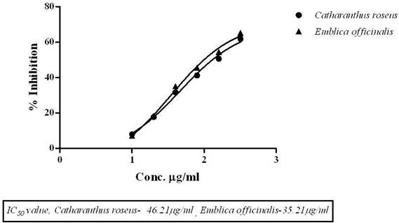
Figure 1. Cytotoxic effect of Catharanthusroseus and Emblicaofficinalison colorectal carcinoma HCT 116 cell line.
References
- Hejmadi M; How cancer arises? In ‘Introduction to cancer biology’, Ed. Hejmadi M Ventus Publishing ApS, United Kingdom, pp 6-12 (2009).
- Tomasetti C, B Vogelstein; Variation in cancer risk among tissues can be explained by the number of stem cell divisions,Science, 347 (6217): 78 (2015). DOI: 10.1126/science.1260825
- Timothy J Y, Colon Cancer; University of South Florida, Encyclopaedia of life Sciences, Nature Publishing Group, 1-2 Tropical Botanical Gardens. (2001). http:/www.els.net
- Gordon M C, J David; Plants as anticancer agents, Journal of ethnopharmacology. 72-75 (2005).
- Wasundara F and H P Vasantha; Anticancer properties of phytochemicals present in Medicinal plants of North America (2012).
- Sumitra C and N Kurnal; In vitro and In vivo methods for anticancer activity evaluation and some Indian medicinal plants possessing anticancer properties: A Review, Journal of Pharmacognosy and Phytochemistry, IC 8192 (2) (2013).
- James R H; Natural Products; the Secondary Metabolites, the Royal Society of Chemistry, UK, (10):1039-40 (2003).
- Pandey Govind; Some important anti-cancer herb- a review, International Research of Pharmacy, 2(7); 45-52 (2011).
- Parimalakrishnan Sundararajanet; Studies of Anticancer and Antipyretic Activity of Bidenspilosa whole plant, African Health Sciences, 6(1):27-30 (2006).
- C Yompakadee, S Thunyaharan; Bacterial activity of methanol extracts of Crabapple, Mangroove Tree against multidrug resistant pathogens, Indian Journal of Pharmaceutical Sciences, 74(3): 230-236 (2012).
- Romijn J C, C F Verkoelen and F H Schroeder; Application of the MTT assay to human prostate cancer cell lines in vitro: Establishment of test conditions and assessment of hormone- stimulated growth and drug induced cytostatic effects. The Prostate, 12(1):99-110 (1988).
- Mebrahtom Gebrelibanos; In-vitro Erythrocyte Haemolysis Inhibition Properties of Sennasingueana Extracts, Momona Ethiopian Journal of Sciences, 4(2): 16-28 (2012).
- Mehboob Hoque, Sandeep Dave, Pawan Gupta, Mohammed Saleemuddin; Oleic Acid May Be the Key Contributor in the BAMLET Induced Erythrocyte Haemolysis and Tumoricidal Action. Plosone: 8(9) 68390(2013).
- Ibrahim I H; Oxidative Haemolysis of Erythrocytes Induced by Various Vitamins, International Journal of Biomedical Sciences, 95-298 (2006).
- Ricardo C B, A L Monica, M G Sonia, M B Fabiana, M A Priscila; A simple method to measure cell viability in proliferation and cytotoxic assays. Braz Oral Res, 23(3): 255-62 (2009).
- Tatsuya Kaneshiro, Masumi Suzui, ReikaTakamatsu, Akira Murakami, Hajime Ohigashi, Tetsuya Fujino, Naoki Yoshimi; Growth Inhibitory Activities of Crude Extracts Obtained from Herbal Plants in the Ryukyu Islands on Several Human Colon Carcinoma Cell Lines, Asian Pacific J Cancer Prev, 6, 353-358 (2005).
- Rafie H, H Soheila, H Mohsen, S Mina; Camphor (Cinnamomum camphora), a traditional remedy with history of treating several diseases. International journal of case reports and images; 4(2): 86-89 (2013).
- Gajalakshmi S, S Vijayalakshmi; Pharmacological activities of Catharanthus roseus: A Perspective Review, International Journal of Pharma and Bio Sciences 4(2): 431 – 439 (2013).
- Ekta S, S Sheel; Phytochemistry; traditional uses and cancer chemopreventive activity of Amla (Phyllanthus emblica): The Sustainer. Journal of Applied Pharmaceutical Science 02 (01); 176-183 (2011).
- Seham M A Moustafa, M Bassem. Menshawi, Gamila M. WASSEL, Khaled Mahmoud, Marwa, M Mounier; Screening of some Plants in Egypt for their Cytotoxicity against four Human Cancer cell lines. International Journal of PharmTech Research; 6 (3): 1074-1084 (2014).
- Irma P, Agnieszka G, S Danuta; Saponins as cytotoxic agents: a review. Phytochem Rev; 9: 425-74 (2010).


