International Journal of Animal Biology, Vol. 1, No. 5, October 2015 Publish Date: Aug. 13, 2015 Pages: 266-272
Curative Effect of Ethanolic Plant Extractives Against Beauveria bassiana Infection in Silkworm, Bombyx mori L.: Histopathological Observations on Midgut
J. A. Chavan1, Y. B. Gaikwad1, A. K. Chougale2, G. P. Bhawane1, *
1Department of Zoology, Shivaji University, Kolhapur, India
2Institute of Science, Fort, Mumbai, India
Abstract
Beauveria bassiana is causative pathogen of white muscardine disease in silkworm Bombyx mori. Occasionally it is responsible for heavy losses to silk industry in many parts of the world. Scrutiny of herbal extracts in the disease management of silkworm, Curcuma longa, Argimone mexicana and Clerodendrum multiflorum showed antifungal activities and these plants were selected for testing their antifungal activities and their doses at 100µl/larva of 6000-ppm concentration were given on calibrated piece of leaf to the B. bassiana inoculated and starved larvae for three consecutive days. Observations on mortality and histopathology of midgut reveals that ethanolic extractive of C. longa has greater potential to check the infection of fungus B. bassiana than the extractive of A. mexicana and C. multiflorum. After making a suitable formulation and evolving farmer friendly strategy of applications, these plant products could be utilized successfully in the management of white muscardine disease in silkworm.
Keywords
Curcuma longa, Argimone mexicana, Clerodendrum multiflorum, White Muscardine
Received: July 10, 2015
Accepted: July 29, 2015
Published online: August 13, 2015
@ 2015 The Authors. Published by American Institute of Science. This Open Access article is under the CC BY-NC license. http://creativecommons.org/licenses/by-nc/4.0/
Contents
1. Introduction 2. Materials and Methods 2.1. Experimental Animals 2.2. Microorganisms 2.3. Antifungal Plants 2.4. Preparation of Plant Extract 2.5. Treatment of Pathogen and Plant Extracts to Silkworm 2.6. Processing of B. mori Tissues for Microscopic Studies 3. Results and Discussion Acknowledgments
1. Introduction
In insect, the midgut is the main site of digestion of food and absorption of digested food besides it known acts as barrier to the variety of parasitic organisms because of chemical composition of gut content, presence of certain non-specific surface inhibitors and the formation of protective peritropic membrane by gut epithelium enclosing the food (Tinsley, 1975). The midgut of B. mori clearly elucidates the columnar cells forming the major portion of the epithelium having striated luminal borders and the goblet cells are intermixed with columnar cells. The epithelium is protected by a peritropic membrane and has regeneration capacity.
White muscardine is the well-known disease of silkworm caused by an entomopathogenic fungus B. bassiana. Bell, (1974) reported this fungus attacked more than 150 insect species. During 1920-25, this disease wiped out the entire sericulture industry in Italy and France. Due to this disease, losses of 10-40% accounted to sericulture industry in Japan and India (Janakiraman, 1961).
Plants are known to contain several classes of secondary metabolite with diverse biological activity. A wide variety of plant secondary metabolites has been identified as active principles for the treatment of various ailments (Taylor et al., 2001; Ncube et al., 2008). Flavonoids constitute a group of important plant secondary metabolites (Subramani et al., 2004). After the discovery of phytoalexin properties of flavonoids and isoflavonoids several investigations have been carried out to evaluate the antimicrobial potential to these compounds (Ramachandraiah and Reddy, 1991; Widenborner and Jha, 1993; Seetharaman et al., 1995; Kuroyanagi et al., 1999 and Kotkar, et al., 2001). Therefore, the present work has been undertaken to know the antifungal effect ethanolic extracts of C. longa, A. mexicana and C. multiflorum on the midgut infection of B. bassiana of mulberry silkworm, B. mori.
2. Materials and Methods
2.1. Experimental Animals
The disease free laying (dfls) of popular pure multivoltine race, Pure Mysore (PM) and bivoltine race, CSR2 was obtained from the Directorate, Government Sericulture Grainage Center, Shahupuri, Kolhapur, for laboratory rearing. The larvae were reared as per the method of Krishnaswamy (1978, 1979) and experiments were conducted during January to August 2011 during which four rearing were done.
2.2. Microorganisms
The fungus culture of B. bassiana was made available from Microbial Type Culture Collection (M.T.C.C), institute of Microbial Technology, Chandigarh, India. The fungus culture was maintained as per the procedure of Govindan et al., (1998).
2.3. Antifungal Plants
For the present work, three plants Viz., C. longa, A. mexicana and C. multiflorum were selected on the basis of earlier reports and by arranging preliminary screening experiments for plant extractives.
2.4. Preparation of Plant Extract
Fresh leaves of C. multiflorum, seeds of A. mexicana and rhizome of C. longa were collected from fields of Kolhapur District, Maharashtra, India. The taxonomic identification of the plants was made with available literature (Yadav and Sardesai, 2002).
The collected plant materials were washed with distilled water and shade dried at room temperature. The materials were grinded to fine powder with the help of mixer grinder. Then these powdered materials were used for preparation of ethanolic extracts by using 50g powder mashed in 100 ml absolute ethanol for 72 hours. The mixture was stirred every 24 hour using a sterile glass rod and it was filtered by using Whatmans filter paper No.1. At the end of extraction, each extract was concentrated in oven at 300C and paste extract was stored at 40C until further use.
2.5. Treatment of Pathogen and Plant Extracts to Silkworm
The B. bassiana was cultured in Petri plates using Sabouraud’s dextrose agar. Conidia were harvested by scrapping the surface of 3 week old culture into a 250 ml glass beaker containing 50 ml sterile distilled water. A drop of tween -20 was then added to the beaker containing distilled water and conidia. The conidial suspension was prepared by mixing the solution using a magnetic stirrer for 5 min and was then diluted in sterilized distilled water to get the desired concentration based on counts made with an improved Neubauer’s Haemocytometer. Nine spore concentration of the fungus Viz., 101 to 109 spores/ml suspension with three replication of each. The experiment was repeated for four rearings. The LC50 was observed at 1x10⁶ spores/ml for PM race and 1x 10⁵ for CSR2 race.
Newly ecdysed fifth instar larvae of PM and CSR2 were topically inoculated by dipping them in the B. bassiana inoculums suspension of 1x10⁶ spores/ml and 1x 10⁵ concentrations respectively. Five groups were arranged control, inoculated and three groups inoculated and separately of plant extracts treated groups. In preliminary study four concentrations viz., 2000 ppm, 4000 ppm, 6000 ppm and 8000 ppm was used for treatment after 6 hours of the inoculation of pathogen, the larvae were fed with ethanolic plant extract in above concentrations of C. longa, A. mexicana and C. multiflorum by spreading 100 µl ethanolic extracts on 4 cm2 piece of mulberry leaf separately and repeatedly for three days in the morning. Normal and inoculated control groups were fed on normal leaves without any application. The survival rate was maximum in 6000 ppm ethanolic extract treated treatments therefore the same was used for further histological studies in midgut of both races.
2.6. Processing of B. mori Tissues for Microscopic Studies
The worms were dissected out in Insect Ringer. The midgut tissues washed in chilled Insect Ringer and then fixed in Bouin’s fixative for 24-48 hrs, dehydrated in the graded alcohol series, cleared in xylene and embedded in paraffin wax. 5 to 7 µm sections were taken, processed for staining with haematoxylene and eosin (HE) (Humason, 1963) and specific fungal staining by Periodic acid Schiff’s (PAS) reaction as per Hotchkiss-McManus method, (McManus, 1948) using fast green as contrast stain.
3. Results and Discussion
The entomopathogenic fungus B. bassiana infects economically important insect silkworm B. mori. It causes the most contagious fungal disease, white musacrdine, and the disease has been responsible for severe cocoon crop loss frequently in all the silkworm rearing areas of the country.
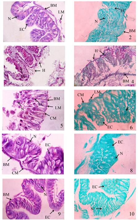
Plate-I. Transverse section of midgut tissue Fig.1and 2. Normal control, 3 and 4. Inoculated control, 5. and 6. C. longa, 7. and 8. A. mexicana, 9. and 10. C. multiflorum treated larvae of PM race.
BM: Basement Membrane
PM: Peritropic Membrane
EC: Epithelial cell
PT: Plant Tissue
H: Hyphae
LM: Longitudinal Muscles
CM: Circular Muscles
N: Nucleus
V: Vacuole
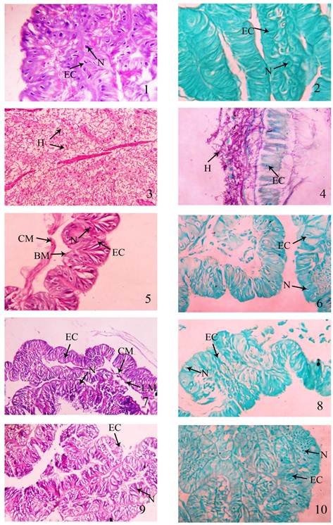
Plate-II. Transverse section of midgut tissue Fig.1 and 2.: Normal control, 3 and 4.: Inoculated control, 5. and 6.: C. longa, 7. and 8.: A. mexicana, 9. and 10.: C. multiflorum treated larvae of CSR2 race.
BM: Basement Membrane
PM: Peritropic Membrane
EC: Epithelial cell
PT: Plant Tissue
H: Hyphae
LM: Longitudinal Muscles
CM: Circular Muscles
N: Nucleus
V: Vacuole
In the fifth instar larvae of silkworm B. mori the midgut represents the largest part of alimentary canal and extended from the 2nd to 9th abdominal segments. The wall of the midgut is composed of outer muscle layer and inner epithelia layer. The midgut epithelium is greatly folded and rests upon a basement membrane. The epithelium of midgut secretes a fine peritropic membrane in the lumen throughout the life of larvae. The midgut was externally covered by circular and longitudinal muscles towards the outer side of the midgut. The epithelial cells and the muscles of the control midgut of PM and CSR2 larvae appear to have normal histological features (Plate-I Fig. 1 and 2, Plate- II Fig. 1 and 2). The cells of midgut epithelium were differentiated into three types- Columnar cells (CC), Goblet cells (GC) and regenerative cells (Mathavan et al., 1989). The CC are tall and closely associated with each other so that their boundaries are indistinct. They are interspersed apically with goblet and basally with RC. The CC possesses a fine brush border facing towards the lumen. The GC is large flask or vase shaped, modified epithelial cells. The GC contains centrally the oval nuclei and bulk of cytoplasm in the basal region. They apically open in the lumen of the midgut. The RC are small and irregular shaped. They are wrapped partially by the basement membrane. These cells can be sharply distinguished from rest of the epithelial cells due to their basal location, small size, centrally located large spherical nuclei with granular cytoplasm.
In case of B. bassiana inoculated larvae the epithelial cells of the entire midgut epithelium were invaded by the fungal mycelia extensively and in the HE stained tissue the disturbed structure of midgut epithelium was observed in both races. When specific fungal staining technique (PAS) was used, in the infected midgut tissue the fungal growth in the tissue stained reddish or pink in colour in both PM and CSR2. The fat bodies, circular and longitudinal muscles of the gut were also get invaded by the hyphae and they stained reddish or pink (Plate-I Fig. 3 and 4, Plate- II Fig. 3 and 4). Almost all the tissues and muscles of the gut were shrunken in size and get compacted lost their original shapes due to fungal manifestation. Results on the similar observed by Jhansi (2000). She observed compactness of the tissues in infected midgut due to pebrine disease.
Kawakami (1973) and Sohaf et al., (1993) were observed penetration of hyphae into the gut, integument and fat bodies. Several researchers reported similar type of effects due to fungal infection in the tissues of the silkworm, B. mori (Mathawan et al., 1991; Aruga, 1994; Kumar et al., 1994; Govindan et al., 1998 and Kumar et al., 1999). The results of the present study about infections are in general agreed with the reports of earlier workers (Krishnaswami, 1987; Jolly, 1987; Ullal and Narasimhanna, 1987; Pringle, 1984).
The ethanolic plant extracts treatement with C. longa, A. mexicana and C. multiflorum after inoculation of fungus showed the significant control (Plate-I Fig. 5, 6, 7, 8, 9 and 10, Plate- II Fig. 5, 6, 7, 8, 9 and 10) because the treatment of these plant extractives within six hours of inoculation probably preventing the establishment of fungal infection in midgut and other tissues of infected silkworm larvae and there was no damage to the midgut epithelium resulting into the survival and subsequent cocooning. The antifungal activities of these plants were reported by (Suresh et al., 1997; Rana et al., 1999 and Khanna, 1999). Barnabas and Nagarajan (1988) reported that, flavonoides extractives prepared from Eucalyptus teriticinis exhibited antifungal and antibacterial activity against several test organism including Aspergillus flavipes and Candida albicum. The secondary metabolites of plants like terpenes and terpenoids are active against fungi reported by several workers (Ayafor et al., 1994; Harrigan et al., 1993; Kubo et al., 1993; Rana et al., 1997; Rao et al., 1993; Suresh et al., 1997; Taylor et al., 1996). The plants secondary metabolites like alkaloids and flavonoids have been known to have antiviral, antibacterial and anticancer agents (Ahn, 1994; Silva et al., 1996; Iwu, 1999; Iwu et al., 1999; Scheck et al., 2006). Two antifungal compounds nimonol and isomeldenin have been isolated from the uncrushed green leaves of Azadirachta indica (Suresh et al., 1997). Manimeghalai and Chandramohan, (2005) used the botanicals Thuja orientalis L. and Curcuma domestica V. inhibiting the growth of Bacillus thuriengensis (flacherie) and in 2006 used Psoralea corylifolia and Plectranthus amboinicus L. for management of grasserie disease of silkworm B. mori.
The results of the present study revealed that the vegetative hyphae penetrated the tissue of the midgut and attacked the epithelial cells in the advanced stage of disease. Midgut tissues get destroyed by the toxins or substances secreted by the fungal hyphae as reported by Yanagita and Iwashita, (1987). The gut epithelium of PM and CSR2 larvae was infected with hyphae of inoculated control group but the treatment of ethanolic plant extracts under study preventing the penetration of hyphae in the midgut tissue of more than 65% of larvae.
Hence, it is concluded that B. bassiana causes the severe damage to the midgut tissue but due to the application of plant extracts having antifungal activity there was no germination and penetration of mycelia in the gut and other tissues and due to which survival rate is 60 to 65% and the silkworms were able to complete their life cycle normally. Therefore, these plant products could be useful to the sericulture industry in the muscardine management after standardizing the doses and making suitable formulation for application.
Acknowledgments
Authors are thankful to UGC, New Delhi for financial assistance and Head Department of Zoology, Shivaji University, Kolhapur for providing laboratory and other infrastructural facilities.
References
- Ahn, J. W. (1994) Cytotoxins limonoids from Melia azedarach var. Japonica. Phytochem., 36: 1493-1496.
- Aruga, H. (1994) Principles of Sericulture, Oxford and IBH Publishing Co PVT Ltd, Tech ED: D. Mahadevappa, Translated by Alamelu Gopal, New Delhi, India.
- Ayafor, J.F.; Tchuendem, M.H.K. and Nyasse, B. (1994)Novel bioactive diterpenoids from Aframomum aulacocarpos. J. Nat. Prod. 57: 917–923.
- Barnabas, C.G. and Nagarajan, S. (1988)Antimicrobial activity of flavonoids of some medicinal plants. Fitoterapia., 3: 508-510.
- Bell, J. V. (1974) Mycoses, In: Insect diseases (ED: Cartwell, GE) Vol-I, Marcel Dekker, Inc, Newyork, 185-236.
- Govindan, R.; Narayanswamy, T. K. and Devaiah, M. C. (1998) Principles of Silkworm Pathology, SERI Scientific Publishers: Bangalore, 31-33.
- Harrigan, G. G.; Ahmad, A.; Baj, N.; Glass, T. E.; Gunatilaka, A. A. L. and Kingston, D. G. I. (1993) Bioactive and other sesquiterpenoids from Porella cordeana. J. Nat. Prod. 56: 921–925.
- Humason, G. L. (1963) Animal tissue techniques.W. H. Freeman and Company.
- Iwu, M. (1999) Garcina cola: A new adaptogen with remarkable immunostimulant, antiinfective and antiinflamatory properties. A colloquium on Garcinia cola. Presented in International Conference of Ethnomedicine and Drug Discovery, Maryland, USA. 26.
- Iwu, M. W.; Duncan, A. R.and Okunji, C. O. (1999) New antimicrobials of plant origin. In: Janick J (editor), Perspectives on new crops and new uses. ASHS press. Alexandria. 457- 462.
- Janakiraman, A. T. (1961) Disease affecting the Indian silkworm races (Rerve ver soic), J. Silkworm., 13: 91-101.
- Jhansi, L. VVNS. (2000) Biochemical changes and diagnosis of microsporodial disease (Pebrine) of silkworm Bombyx mori L. (Lepidoptera: Bombycidae). J. Ent. Res., 24(4): 301-310.
- Jolly, M. S. (1987) Appropriate Sericulture Techniques, Published by Director, International Centre for Training and Research in Tropical Sericulture, Mysore, India.
- Kadway, M. (2009) Studies on the effect of pathogens on some vital organs of tropical tasar silkworm, Antheria mylita (Drury) (Lepidoptera: Noctuidae). Ph. D. thesis submittedto RTM, Nagpur University, Nagpur.
- Kawakami, K. (1973) Studies on the muscardine disease of the silkworm Bombyx mori L with special reference to the invasion of causative fungi and pathological changes in infected larva, Bull Sericul Exp Sta, 25 (5): 369-370.
- Khanna, N. M. (1999) Turmeric-Natures precious gift. Current Science, 76: 10: 1351-1356.
- Kotkar, H. M.; Mendaki, P. S.; Sadan, S. V. G. S.; Jha, S. R.; Upasani, S. M., and Maheshwari, V. L. (2001) Antimicrobial and pesticides activity of partially purified flavonoides from Annona squamossa. Pest Manag. Sci. 58: 33-37.
- Krishanaswami, S. (1978) New technology of silkworm rearing Central Sericulture Research and Training Institute, Mysore, Bull. 3 (2): 1-23.
- Krishnaswami, S. (1979) Improved method of rearing young age silkworm, C. S. R and TI Bulletin, No. 3: 1-24.
- Krishnaswami, S.; Narasimhanna, M. N.; Suryanarayana, S. K. and Kumararaj, S. (1987) Manual on Silkworm Rearing, Sericulture – Vol 2, FAO Agri Service Bull, 15/2, Central Silk Board, Bangalore, Rome.
- Kubo, I.; Muroi, H. and Himejima, M. (1993) Combination effects of antifungal nagilactones against Candida albicans and two other fungi with phenylpropanoids. J. Nat. Prod. 56: 220–226.
- Kumar, V.; Singh, G. H.; Basui, A. M.; Ahsam, M. M. and Datta, R. K. (1999) Germination, Penetration and inavasion of Beauveria bassiana causing white muscardine, the Italian Journal of Zoology., 66(1): 10-14.
- Kumar, V.; Tewari, S. K. and Awasthi, A. K. (1994) Surface ultrastructure of Beauveria bassiana infecting silkworm Bombyx mori L, Current Science., 67(7) : 546-548.
- Kuroyanagi, M., Arakawa, T., Hirayama, Y. and Hayashi, T., 1999. Antibacterial and antiandrogen flavonoids from Sophora flavescens. J. Nat. Prod., 62: 1595-1599.
- Manimeghalai, S. and Chandramohan, N. (2005) Botanicals for the management of Bacterial flacheri of silkworm, Bombyx mori L. Sericologia., 45(1): 51-58.
- Manimeghalai, S. and Chandramohan, N. (2006) Efficacy of botanicals against grasserie disease of silkworm, Bombyx mori L. Sericologia., 46(1): 15-23.
- Mathavan, S.; Sudha, P. M. and Muthu, S. P. (1991) Histological and histopathological studies on midgut epithelium of Bombyx mori larvae affected by Bacillus sphaericus. Sericologia., 31(3): 403-411.
- Mcmanus, J. F. A. (1948) Histological and histochemical uses of periodic acid. Stain technology. 23: 99-108.
- Ncube, N. S.; Afolayan, A. J. and Okoh, A. I. (2008) Assessment techniques of antimicrobial properties of natural compounds of plant origin: current methods and future trends. Afr. J. Biotchnol. 7: 1797-1806.
- Pringle, J. A. (1984) Report on the diseases of silkworms in India, International Books and Periodicals supply services, 24B/5, Deshbandhu gupta road, Karol Bagh, New Delhi., 57-60.
- Ramachandraiah, P. and Reddy, M. B. (1991) Antifungal activity of some naturally occurring flavonoids. Indian J. Microbiol., 31: 55-56.
- Rana, B. K.; Singh, U. P. and Taneja, V. (1999) Antifungal activity and kinetics of inhibition by essential oil isolated from leaves of Aegle marmelos. J Ethnopharmacol., 57: 29-30.
- Rao, K. V.; Sreeramulu, K.; Gunasekar, D. and Ramesh, D. (1993) Two new sesquiterpene lactones from Ceiba pentandra. J. Nat. Prod. 56: 2041–2045.
- Scheck, A. C.; Perry, k.; Hank, N. C. and Clark, W. D. (2006) Anticancer activity of extracts derived from the mature roots of Scutellaria baicalensis on human malignant brain tumor cells. Biomed. Centr. Complement. Alternat. Med. 6-27.
- Seetharaman, T. R.; Thirunarayanan, S. and Ganesan, T. (1995) Antifungal activity of flavonol glycosides. Geobios., 22: 87-89.
- Shoaf, K. A.; Chisti, M. Z. and Trag, A. R. (1993) Histopathology of the silkworm Bombyx mori L infected with Beauveria bassiana (Bals) Vuill, Indian. J. Seric., 32(2): 213-215.
- Silva, O.; Duarte, A.; Cabrita, J.; Pimentel, M.; Diniz, A. and Gomez, E. (1996) Antimicrobial activity of Guinea-Bissau traditional remedies. J. Ethnopharmacol. 50: 53-59.
- Subramani, K.; Gunasegaran, R.; Shiyamala, C. and Ganesan, T. (2004)Antifungal activity of Certain flavonol glycosides and phenolic acid from Canthium Species. Ad. Plant. Sci., 17(11): 745-748.
- Suresh, B.; Sriram, S.; Dhanaraj, S. A.; Elango, K. and Chinnaswamy, K. (1997) Anticandidal activity of Santolina chamaecyparissus volatile oil. J. Ethnopharmacol. 55: 151–159
- Suresh, G.; Narasimhan, N. S.; Masilamani, S.; Partho, P. D. and Gopalkrishnan, G. (1997) Antifungal fractions and compounds from uncrushed green leaves of Azadirachta indica. Phytoparasitica. 25(1): 33-39.
- Taylor, J. L. S.; Robe, T.; Mcgaw, L. J.; Jager A. k. and Van, S. J. (2001) Towards the scientific validation of traditional medicinal plants. Plant Growth Regl., 34: 23-37.
- Taylor, R. S. L.; Edel, F.;Manandhar, N. P. and Towers, G. H. N. (1996) Antimicrobial activities of southern Nepalese medicinal plants. J. Ethnopharmacol., 50: 97–102.
- Tinsley, T. W. (1975) Factors affecting virus infection of insect gut tissue. In "Invertebrate immunity" (K. Maramorosch and R. E. Shope, eds.), pp. 55-63. Academic press, New York.
- Ullal, S. R. and Narasimhanna, M. N. (1987) Handbook of Practical Sericulture, Central Silk Board (Ministry of Textiles-Government of India), United Mansion, 39, Mahatma Gandhi Road, Bangalore, 138-151.
- Widenborner, M. and Jha, H. C. (1993) Antifungal activity of flavonoids and their mixtures against different fungi occurring on gain. Pestic. Sci., 38: 347-351.
- Yadav, S. R. and Sardesai, M. M. (2002) Flora of Kolhapur District, Shivaji Univerity Publication., pp. 371.
- Yanagita, T. and Iwashita, Y. (1987) Histological observation of larvae of the silkworm, Bombyx mori orally infected with Beauveria bassiana, J. Seric. Sci, Jpn., 56(4): 285.


