International Journal of Animal Biology, Vol. 1, No. 4, August 2015 Publish Date: Jul. 9, 2015 Pages: 99-105
Effect of Vernonia amygdalina on Some Biochemical Indices in Dimethylnitrosamine (DMN)-Induced Liver Injury in Rats
Usunobun Usunomena1, *, Okolie P. Ngozi2, Eze G. Ikechi3
1Department of Basic Sciences (Biochemistry Unit), Faculty of Basic and Applied Sciences, Benson Idahosa University, Benin City, Edo State, Nigeria
2Department of Biochemistry, Faculty of Life Sciences, University of Benin, Benin City, Nigeria
3Department of Anatomy, School of Basic Medical Sciences, College of Medicine, University of Benin, Benin City, Nigeria
Abstract
This experiment pertains to the protective role of Vernonia amygdalina pre-treatment on some biochemical indices in dimethylnitrosamine (DMN)-induced liver damage in male albino rats. Four (4) groups of six (6) rats each were used for the study. Group 1served as control, group 2 and 3 were pre-treated with 400mg/kg Vernonia amygdalina for one week while group 3 and 4 each received single dose of 20mg/kg DMN (orally) after one week. The rats were sacrificed 48hrs after DMN administration. In rats administered 20mg/kg DMN, liver damage was clearly shown by increased activities of serum hepatic marker enzymes namely aspartate aminotransaminase (AST), alanine aminotransaminase (ALT), alkaline phosphatase (ALP), and gamma glutamyltransferase (GGT), increased lipid profile parameters such as total cholesterol and triglycerides as well as increased level of lipid peroxidation indices, malondialdehyde (MDA) in liver. The toxic effect of DMN was also indicated by significantly decreased levels of antioxidants such as superoxide dismutase (SOD), catalase (CAT) and reduced glutathione (GSH). However, in rats pre-treated with 400mg/kg Vernonia amygdalina and dosed thereafter with DMN, there were significant reversal in the activities of serum hepatic marker enzymes, lipid profiles, lipid peroxidation and significant restoration of antioxidant levels in the liver when compared to DMN-alone treated rats. Histopathological studies in the liver of rats also showed that Vernonia amygdalina pre-treatment markedly reduced the toxicity of DMN and significantly preserved the normal histological architecture of the tissue. The findings of this study suggest that pre-treatment with Vernonia amygdalina leaves has a protective and beneficial effect on liver subjected to DMN-induced oxidative stress, possibly by decreasing lipid peroxidation and enhancing endogenous antioxidant production.
Keywords
Antioxidant, Dimethylnitrosamine, Necrosis, Oxidative Stress, Vernonia Amygdalina
Received: June 15, 2015
Accepted: June 17, 2015
Published online: July 7, 2015
@ 2015 The Authors. Published by American Institute of Science. This Open Access article is under the CC BY-NC license. http://creativecommons.org/licenses/by-nc/4.0/
Contents
1. Introduction 2. Materials and Methods 2.1. Collection, Identification, Preparation and Extraction of Plant Leaves 2.2. Experimental Animals, Dmn and Extract Administration 2.3. Collection of Tissue Samples and Preparation of Liver Homogenates 2.4. Biochemical Assays 2.5. Histology 3. Results 4. Discussion
1. Introduction
Vernonia amygdalina, also known as ‘‘African bitterleaf’’, is a plant vegetable used for both food and traditional treatment of diseases throughout tropical Africa (1). Nutritional and phytochemical evaluations have revealed high levels of antioxidant vitamins, mineral elements (Fe, Se, Zn,Cu, Cr and Mn) and phyto-compounds (flavonoids, saponins, alkaloids and tannins) in the leaves of Vernonia amygdalina (2-4). The aqueous leaf extract have been shown to posses anti-hyperlipidemic and hypolipidemic effect respectively on diabetic and non-diabetic rats (5). Its protective role on the kidneys (6) of alloxan-diabetic rats has additionally been investigated and results reported. The plant has acquired special relevance, having been shown in human medicine to possess potent anti-tumorigenic properties (7) with an amazing anti-parasitic efficacy in zoo pharmacognosy, as it is easily recognized and used for self medication by parasitized chimpanzees (8).
Dimetylnitrosamine (DMN) is a potent hepatotoxin, which is metabolized by microsomal cytochrome p450IIE1 in liver (9). Activation of DMN by CYP2E1 in mouse liver has been shown to stimulate Kupffer cells leading to generation of superoxide and other reactive oxygen species (ROS) capable of damaging liver cells (10). The occurrence of DMN in our drinks and foods including fish, meat as well as fresh supermarket products is well established (11-14). In a food survey conducted by Domanska and Kowalski (15), DMN was detected in 31.5% of analyzed samples with the major dietary sources being cooked meat products, cooked fish and spices. Proksch (16) reported the presence of DMN in rubber products including gloves, balloons, toys, baby bottle teats, soothers and condoms. This study is aimed at investigating the ability of ethanolic leaf extract of Vernonia amygdalina pre-treatment to protect the liver against dimethylnitrosamine (DMN)-induced hepatocellular damage and oxidative stress in rats in vivo.
2. Materials and Methods
2.1. Collection, Identification, Preparation and Extraction of Plant Leaves
Fresh leaves of Vernonia amygdalina were purchased from a local market in Benin City, Edo state, Nigeria. The leaves were identified by Dr. Chris Akoma, a Botanist in the Department of Basic Sciences, Faculty of Basic and Applied Sciences, Benson Idahosa University, Benin city, Edo State. The Vernonia amygdalina leaves were separated from the stalk, washed and air-dried at room temperature (24ᵒC) and then pulverized, crushed into fine powder and weighed.
Ethanolic extracts of the plant leaves was prepared by soaking 250g of the dry powdered plant leaves in one (1) litre of absolute ethanol at room temperature for 48hrs. The extract was then filtered first through a Whatmann filter paper No. 42 (125mm) and then through cotton wool. The extract was thereafter concentrated using a rotary evaporator with the water bath set at 40ºC to one-tenth its original volume and then finally freeze dried. The dried residue (crude extract) was then stored at 4ºC. Aliquot portions of the crude plant extract residue were weighed and dissolved in distilled water for use on each day of our experiments.
2.2. Experimental Animals, Dmn and Extract Administration
Male wistar albino rats divided into seven groups of six (6) rats each, weighing between 160-195g were obtained from the Animal Unit facility of the University of Ibadan, Oyo state, Nigeria and housed in wooden cages in the animal house of the Department of Biochemistry, University of Benin. The rats were maintained under controlled environmental conditions (temperature—24±2◦C;relative humidity—50–70%; 12 h light/dark cycle), housed for one week after their arrival to the animal house for acclimatization. The rats had free access to drinking water and normal pellet diet (NPD) ad libitum until they were assigned to individual groups. Institutional Animal Ethical Committee permission was obtained before performing the experiments.
DMN used in this work was synthesized in a fume chamber at the Department of Biochemistry, University of Ibadan, Oyo state, Nigeria, according to the method of Vogel (17).
A total of 24 rats divided into 4 groups were used. Group 1 served as control and was given normal saline, Group 2 received 400mg/kg Vernonia amygdalina only for seven (7) days consecutively, Group 3 received 400mg/kg Vernonia amygdalina for 7 days consecutively followed by oral administration of a single dose of 20mg/kg DMN (dissolved in 0.15MNaCl), on day 8 (48 hours before sacrifice) while Group 4 received oral administration of single dose of 20mg/kg DMN (dissolved in 0.15MNaCl) on day 8.Before use, the Vernonia amygdalina leaf extract was reconstituted in distilled water and administered orally via gastric intubation. All rats were sacrificed on the tenth day of the study by cardiac puncture and blood collected via the ocular vein in plain tubes and allowed to stand for 45 min before it was centrifuged at 4,000 rpm for 30 min. Serum was stored at -20º C until analyzed.
2.3. Collection of Tissue Samples and Preparation of Liver Homogenates
Following sacrifice, liver samples were quickly excised and rinsed with normal saline. A small portion of each liver sample was fixed in 10% phosphate-buffered formalin for histological examination while the remaining portions were stored at –20°C for biochemical analysis. 10% liver homogenate was prepared in physiological saline. The homogenate was centrifuged at 5000 x g for 15 minutes and the clear supernatant obtained used for biochemical analysis.
2.4. Biochemical Assays
Serum AST and ALT activities were estimated colorimetrically according to the method of Reitman and Frankel (18), ALP, γ-GT, total cholesterol and triglyceride assay were carried out using Randox kits (UK) according to manufacturer’s instructions. GSH was estimated colorimetrically by measuring the reduction of Ellman’s reagent (5, 5’di-thio-bis-2-nitrobenzoic acid) at 412nm as described by Ellman (19). SOD was assayed based on the ability of the enzyme to inhibit the autooxidation of epinephrine according to the method of Misra and Fridovich (20). The assay of CAT was carried out colorimetrically based on the measurement of the rate of decomposition of H2O2 after the addition of the sample containing the enzyme by reacting it with excess KMnO4 and then measuring the residual KMnO4 spectrophotometrically at 480nm (21). MDA was estimated in a colorimetric reaction with thiobarbituric acid (22).
2.5. Histology
Liver sections fixed in formol-saline were processed for light microscopy at the Department of Anatomy, Faculty of Basis Medical Sciences, College of Medicine, University of Benin. The resultant slides were read and interpreted by one of us, G.I.E., a consultant pathologist.
3. Results
Tables 1 illustrate the effect of Vernonia amydalina pre-treatment on the activity of AST, ALT, ALP and GGT in the serum of DMN-administered rats. The levels of AST, ALT, ALP and GGT were significantly (P < 0.05) increased in DMN-administered rats when compared to the control normal and extract treated rats. Administration of Vernonia amygdalina to DMN-administered rats restored all these changes to near normal levels by significant (P < 0.05) reduction of the activity of AST, ALT, ALP and GGT.
Table 1. Effect of ethanolic leaf extracts of Vernonia amygdalina pre-treatment on serum Liver function enzymes in DMN toxicity.
| Treatment | AST (U/l) | ALT (U/l) | ALP (U/l) | GGT (U/l) |
| Control (normal saline) | 19.11±2.28a | 14.80±1.46a | 26.17±2.05a | 13.11±1.31a |
| VAE alone (400mg/kg) | 18.00±3.16a | 12.50±3.57a | 23.33±0.94a | 11.87±2.43a |
| VAE (400mg/kg) + DMN (20mg/kg) | 120.75±2.75b | 83.75±3.56b | 44.10±1.63b | 28.12±2.00b |
| DMN alone (20mg/kg) | 201.00±7.47c | 138.25±6.26c | 92.67±5.26c | 51.00±3.12c |
Values are expressed as Mean ± SD, (n=5), VAE = Vernonia amygdalina, ADMN = Dimethylnitrosamine, AST = Aspartate aminotransferase, ALT = Alanine aminotransferase, ALP = Alkaline phosphatase, GGT = Gamma-glutamyltransferase
Mean values in each column having different superscript (a, b, c) are significantly different (p ˂ 0.05) while mean values with same superscript is not significantly different (p ˂ 0.05)
Serum total cholesterol and triglyceride in the ‘control’, DMN-treated, VAE + DMN-treated, and VAE-treated rats are shown in Tables 2. Serum total cholesterol and triglycerides were significantly elevated (P<0.05) in DMN-treated Group D rats as compared to ‘control’ Group A rats. The lipid parameters examined were improved towards normal values following Vernonia amygdalina pre-treatment in Group C rats.
Table 2. Effect of ethanolic leaf extracts of Vernonia amygdalina pre-treatment on serum Total cholesterol and Triglyceride levels in DMN toxicity.
| Treatment | Triglyceride (mg/dl) | Total Cholesterol (mg/dl) |
| Control (normal saline) | 94.12 ± 3.64a | 91.09 ± 8.45a |
| VAE alone (400mg/kg) | 79.00 ± 17.52a | 87.75 ± 10.96a |
| VAE (400mg/kg) + DMN (20mg/kg) | 106.50 ± 3.29b | 133.25 ± 8.88b |
| DMN alone (20mg/kg) | 145.50 ± 5.03c | 168.55 ± 5.03c |
Values are expressed as Mean ± SD, (n=5), VAE = Vernonia amygdalina, DMN = Dimethylnitrosamine.
Mean values in each column having different superscript (a, b, c, bb) are significantly different (P ˂ 0.05) while mean values with same superscript is not significantly different (P ˂ 0.05)
Table 3shows the effects of Vernonia amygdalina ethanolic leaf extract pre-treatment on oxidative stress variables in DMN-administered animals. The hepatic antioxidant activities of CAT, SOD and GSH were significantly decreased (P<0.05), while MDA was significantly increased in the DMN-administered rats. The ‘control’ group of rats maintained optimal values of the antioxidants studied. Vernonia amygdalina pre-treatment significantly (P<0.05) decreased DMN-administered elevated MDA and also significantly increased (P<0.05) DMN-administered reduced antioxidant enzyme activities (CAT, SOD and GSH).
Table 3. Effect of ethanolic leaf extracts of Vernonia amygdalina on Oxidative stress parameters in DMN toxicity.
| Treatment | MDA (U/mg wet tissue) | GSH (µM/mg tissue) | SOD (U/mg wet tissue) | CAT (U/mg wet tissue) |
| Control (normal saline) | 2.53 ± 0.17a | 51.03 ± 5.11a | 11.13 ± 0.74a | 50.34 ± 3.01a |
| VAE alone (400mg/kg) | 2.16 ± 0.10z | 63.09 ± 4.76z | 14.08 ± 1.04z | 60.05 ± 1.40z |
| VAE (400mg/kg) + DMN (20mg/kg) | 4.01 ± 0.04b | 38.17 ± 2.65b | 7.01 ± 0.33b | 37.30 ± 2.03b |
| DMN alone (20mg/kg) | 6.99 ± 0.18c | 21.32 ± 2.18c | 4.45 ± 0.13c | 22.49 ± 1.89c |
Values are expressed as Mean ± SD, (n=5), VAE = Vernonia amygdalina, DMN = Dimethylnitrosamine, MDA=Malondialdehyde, GSH=Reduced glutathione, SOD=Superoxide dismutase, CAT=Catalase
Mean values in each column having different superscript (a, b, c, d, z) are significantly different (p ˂ 0.05) while mean values with same superscript is not significantly different (p ˂ 0.05).
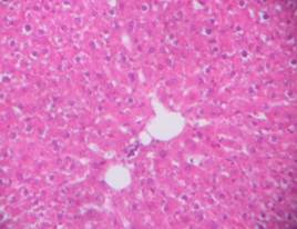
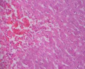
Photomicrographs of liver showed the normal structure of control rats (A). Rats treated with DMN resulted in severe hemorrhagic centrilobular necrosis along with disarrangement of hepatic strands (D). Vernonia amygdalina pre-treatment on acute DMN administration brought back the cellular arrangement around the central vein and reduced necrosis (C).
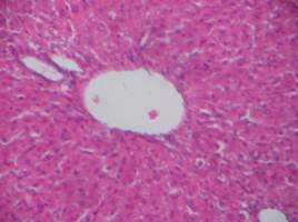
Photomicrograph A. Control rat liver composed of portal vein, hepatocytes separated by sinusoids (H&E x 100).
Photomicrograph B. Rat liver pre-treated with Vernonia amygdalina (400mg/kg) only for 7 days showing fairly unremarkable hepatocytes and mild sinusoidal congestion (H&E x 100).
Photomicrograph C. Rat liver pre-treated with 400mg/kg Vernonia amygdalina for 7 days followed by 20mg/kg DMN on day 8, showing focal areas of haemorrhagic necrosis and viable hepatocytes (H&E x 100).
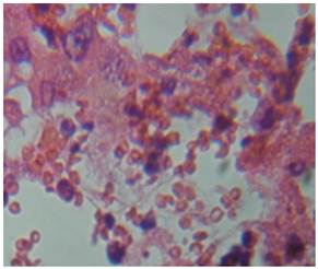
Photomicrograph D. Rat Liver treated with 20mg/kg DMN, 48hours before sacrifice showing severe centrilobular. haemorrhage and necrosis.
4. Discussion
AST, ALT and ALP are enzymes present in higher concentrations in cytoplasm, but when there is hepatopathy, these enzymes leak into blood stream in conformity with the extent of liver damage (23).GGT and ALP are membrane-bound enzymes, which are released unequally depending on the pathological phenomenon. In this study, rats treated with DMN alone showed a significant increase in serum levels of AST, ALT, ALP and GGT compared to controls, thus indicating damage to hepatic cells. Also, alterations in GGT and ALP are likely to affect membrane permeability and produce derangement in the transport of metabolites.
However, DMN-administered rats orally pre-treated with extracts of 400mg/kg Vernonia amygdalina showed significantly decreased levels of serum AST, ALT, ALP and GGT compared to DMN alone treated rats. The decrease in AST, ALT, ALP and GGT levels by the extracts in tested groups, indicates the protection of structural integrity of hepatocytic cell membrane or regeneration of damaged liver cells. Our result is in agreement with earlier observations of Babalola et al (24) that pre-treatment with sesquiterpene lactone extract from the leaf of Vernonia amygdalinaa meliorates CCl4-induced hepatotoxicity in rats. Moreover, Iwalokunet al (25) reported a dose dependent reversal of acetaminophen-induced alterations in mouse liver enzymes by pre-treatment of Vernonia amygdalina. In a related study by Adesanoye and Farombi (26) and Arhoghro et al (27), administration of Vernonia amygdalina resulted in accelerated reversion of hepatic damage caused by CCl4 via reduction of liver marker enzymes like ALT, AST, ALP and bilirubin. The efficacy of any hepatoprotective drug is dependent on its capacity of either reducing the harmful effect or restoring the normal hepatic physiology that has been disturbed by a hepatotoxin. The extracts thus protected the hepatocytes from DMN-induced injuries.
Alterations in the concentration of major lipids of animals such as cholesterol and triglycerides can give useful information on lipid metabolism as well as predisposition of the animals to cardiovascular risk (28).The results of this study shows that DMN caused a significant (P<0.05) increase in the levels of total cholesterol and triglycerides. The rise in serum total cholesterol and triglycerides may also be attributed to increased lipolysis, mediated by increased norepinephrine release which act through interference with the intracellular functions of Ca2+ in the cytoplasm, events that may lead to increased production of ROS, inducing oxidative stress resulting in metabolic dysfunction (29, 30).It is also possible that since LDL and VLDL are produced and degraded in the liver (31), DMN-induced damage may have affected the capacity of the liver to degrade LDL and VLDL leading to hypercholesterolemia. The significant increase in serum total cholesterol and triglycerides in DMN intoxicated group is in agreement with those of Ismail et al (32) who reported that injection with CCl4 increased serum and tissue lipid profile.
However, pre-treatment with extract of 400mg/kg Vernonia amygdalina on acute DMN toxicity showed a significant (P < 0.05) decrease in serum total cholesterol and triglyceride levels. Extracts pre-treatment prior to DMN-administration caused significant reduction in serum total cholesterol, probably due to marked reduction of VLDL cholesterol. The lipid lowering effects of the extracts might also be attributed to an inhibitory activity on acyl-CoA: cholesterol acyltransferase in vivo. This enzyme is responsible for acylation of cholesterol to cholesterol esters in liver (33). Aqueous leaf extract of Vernonia amygdalina have been shown to have hypolipidemic effect in diabetic rats (34) and could be related to the presence of alkaloids, saponins, flavonoids and polyphenols known to reduce serum lipid level in animals (35).
Oxidative stress has been implicated as a factor that contributes to various forms of cell death as free radicals react with lipids causing peroxidation, resulting in the release of products such as malondialdehyde, hydrogen peroxide, and hydroxyl radicals (36).This study shows that DMN caused a decrease in intracellular GSH, SOD and CAT level with concomitant increase in MDA levels in DMN-treated rats compared to controls. There was clear evidence that DMN-induced hepatic injury was associated with free radical injury and oxidative stress. Oxidative stress was characterized by increased lipid peroxidation and/or altered non-enzymatic and enzymatic antioxidant systems. The increase in MDA indicates increased oxidative damage to cell membranes, inhibition of several important enzymes, reduced cellular function, and cell death (36).The observed decrease in hepatic GSH could be due to decreased synthesis, or increased degradation of GSH by DMN-induced oxidative stress.
However, Vernonia amygdalina pre-treatment prior to DMN administration showed increase in GSH, CAT and SOD as well as significant reduction in MDA levels compared to DMN alone treated rats. The reduced MDA may be related to the antioxidant properties of the phytochemical compounds found in the extracts as compounds such as flavonoids and tannins have been reported to exert antioxidant activity by scavenging free radicals that cause lipid peroxidation (37, 4, 38).In a related study by Kujawska et al (39), CCl4 caused significant GSH depletion soon after administration, thus, making glutathione a critical determinant of tissues susceptibility to oxidative damage.
Histopathological analysis shows that when the rats were treated with DMN alone, the liver exhibited massive and severe haemorrhagic necrosis at the centrilobular zone as well as severe vacuolation of hepatocytes similar to previous results (40-42).
However, Vernonia amygdalina pre-treatment prior to DMN administration mitigated the above histopathological changes as the integrity of the hepatocytes were relatively well preserved by inhibiting further tissue necrosis and inflammatory cell infiltration. In a related study, Farombi et al (43, 40) reported that pre-administration of kolaviron and curcumin to DMN treated rats enhanced the hepatocytes integrity as they were relatively preserved.
A number of earlier investigators have shown that tannin and other polyphenolic compounds (e.g., coumarins), flavonoids, triterpenoid, saponins, and a host of other plant secondary metabolites possess hypoglycaemic, hypolipidaemic, hypotensive, anti-inflammatory, and other pharmacological and biochemical properties in various experimental animal models (44). In our previous study, we reported the presence of flavonoids, triterpenoids, tannins, saponins, ascorbic acid and alkaloids in Vernonia amygdalina leaves (4, 37). We can thus speculate that some of the above chemical constituents of Vernonia amygdalina leaves, especially the flavonoids, tannins, saponins and triterpenoids, are probably responsible for the altered biochemical variables in the hepatic tissues, as well as the anti-inflammatory property of Vernonia amygdalina observed in this study.
References
- Farombi, E. O., and Owoeye, O. (2011). Antioxidant and chemopreventive properties of Vernonia amygdalina and Garcinia biflavonoid. International Journal of Environmental Research and Public Health 8: 2533–2555.
- Igile, G. O., Oleszek, W., Jurzysta, M., Burda, S., Fanfunso, M., and Fasanmade, A. A. (1994). Flavonoids from Vernonia amygdalina and their antioxidant activities.Journal of Agricultural and Food Chemistry 42: 2445–2448.
- Atangwho, I. J., Ebong, P. E., Eyong, E. U., Williams, I. O., Eteng, M. U., & Egbung, G. E. (2009). Comparative chemical composition of leaves of some antidiabetic medicinal plants: Azadirachta indica, Vernonia amygdalina and Gongronema latifolium. African Journal of Biotechnology, 8(18): 4685–4689.
- Usunobun U and Okolie N. P (2015). Phytochemical, trace and mineral composition of Vernonia amygdalina leaves. International Journal of Biological & Pharmaceutical Research. 6(5): 393-399.
- Atangwho I. J, Ebong P.E, Egbung G.E, Eteng M.U and Eyong E.U (2007a) Effect of Vernonia amygdalina Del. on liver function in alloxan-induced hyperglycaemic rats. Journal of Pharmacy and Bioresources4(1): 25-31
- Atangwho I. J, Ebong P. E, Eteng M.U, Eyong E.U and Obi A.U (2007b). Effect of Vernonia amygdalinadel leaf on kidney function of diabetic rats. Int. J. Pharmacol., 3: 143-148.
- Izevbige E.B, Bryant T.L and Walker A. (2004). A novel natural inhibitor of extracellular signal regulated kinases and human breast cancer cell growth.Experimental Biol. Med. 229(2): 163-169.
- Huffman, M.A (2003). Animal self-medication and ethno-medicine: Exploration and exploitation of medicinal properties of plants. Proceedings of the Nutrition Society, 62:371-381.
- Guengerich, F.P., D.H. Kim and M. Iwasaki, (1991). Role of human cytochrome P-450 IIE1 in the oxidation of many low molecular weight cancer suspects.Chem. Res. Toxicol., 4(2): 168-179.
- Teufelhofer, O., W. Parzefall, E. Kainzbauer, F. Ferk, C. Freiler, S. Knasmuller, L. Elbling, R. Thurman and R. Schulte-Hermann, (2005). Superoxide generation from Kupffer cells contributes to hepatocarcinogenesis: Studies on NADPH oxidase knockout mice. Carcinogenesis 26: 319-329.
- Okafor P. N. and Ogbonna, U.I (2003). Nitrate and nitrite contamination of water sources and fruit juices marketed in South-Eastern Nigeria. J. Food. Comp. Anal. 16: 213-216.
- Okafor P. N. and Nwaogbo E (2005). Determination of nitrate, nitrite and N-nitrosamines, cyanide and ascorbic acid content of fruit juices marketed in Nigeria. Afri. J. Biotech. 4(10): 1105-1108.
- Okafor, P.N., O. Nwosu, J. Chukwu, J. Agbayi and E.N. Maduagwu, (2007). Occurrence of malondialdehyde and N- nitrosamines and their precursors in some Nigerian ice creams, yogurts, meat and fish species.Afr. J. Biochem. Res., 1(1): 001-005.
- Griesenbeck, J.S., D.S. Michelle, C.H. John, R.S. Joseph, A.R. Antonio and D.B. Jean, (2009). Development of estimates of dietary nitrates, nitrites and nitrosamines for use with the short willet food frequency questionnaire.Nutr. J., 8(16): 1-9.
- Domanska, K. and B. Kowalski, (2002). Effect of different storage conditions on N-nitrosamine content in Polish edible offals processed meat products. Bull. Ver. Inst. Pulawy 46: 317-324.
- Proksch, E., 2001. Toxicological evaluation of nitrosamines in condoms. Int. J. Hyg. Environ.Publishers: New York.
- Vogel. A. I. (1971). A textbook of practical organic Chemistry including qualitative organic analysis. Longman group limited, London.Pp 426.
- Reitman S. and Frankel, S. (1957). A colorimetric method for the determination of SGOT and SGPT. Am. J. Cln. Path.28: 56-58.
- Ellman, G.L. (1959). Tissue sulfhydryl groups.Arch. Biochem. Biophys. 82: 70–77.
- Misra, H. P. and Fridovich (1972). The role of superoxide anion in the autooxidation of epinephrine and a simple assay of superoxide dismutase. J. Biol. Chem., 247: 3170-3175.
- Cohen, G., Dembiee, D. and Marcus, J. (1970). Measuremment of catalase activity in tissues extracts. Anal.Biochem., 34: 30-38.
- Ohkawa H, Ohishi N, and Yogi K, (1979). Assay for lipid peroxidation in animal tissues by thiobarbituric acid reaction. Annals of Biochemistry 95: 351–358.
- Venukumar M.R., and Latha M.S. (2002). Antioxidant activity of Curculigoorchioides in carbon tetra chloride induced hepatopathy in rats. Indian J.Clin.Biochem.17:80-87.
- Babalola O.O., Anetor J.I., and Adeniyi F.A. (2001).Amelioration of carbon tetrachloride induced hepatotoxicity of terpenoid extract from leaves ofVernonia amygdalina.Afr. J. Med. Sci.30(1-2): 91-93.
- Iwalokun, B. A.; Efedede, B. U.; Alabi-Sofunde, J. A.; Oduala, T.; Magbagbeola, O. A.; Akinwande, A. I.(2006).Hepatoprotective and antioxidant activities of Vernonia amygdalina on acetaminophen-induced hepatic damage on mice. Journal of Medicinal Food 9(4):524-30.
- Adesanoye O.A., and Farombi E.O. (2009). Hepatoprotective effects of Vernonia amygdalina (astereaceae) in rats treated with carbon tetrachloride. Exp. Toxicol. Pathol. 62: 197-206.
- Arhoghro E.M., Ekpo K.E., Anosike E.O., and Ibeh G.O. (2009). Effect of aqueous extract of bitter leaf (Vernonia amygdalina Del) on carbon-tetrachloride (CCl4) induced liver damage in albino wistar rats. Eur. J. sci. Res., 26:122-130
- Yakubu M.T., Akanji M.A., and Oladiji A.T. (2008). Alterations in serum lipid profile of male rats by oral administration of aqueous extract of Fadogia argrestis stem. Res. J. Med. Plant. 2: 66-73.
- Gesquieve L, Loreau N, Minnich J. (1999). Oxidative stress leads to cholesterol accumulation in vascular smooth muscle cells. Free Radic Bio Med.27: 134–145.
- Sharma V. and Singh M.(2012). Alterations induced by n-nitrosodimethylamine and ethanolic root extract of Operculina turpethumin serum lipid profile of male albino mice. Asian J. Pharm.Clin. Res., 5(3): 69-73
- Gillian-Daniel L. Donald, Paul W. Bates, Angie Tebon, and Alan D. Attie (2002). Endoplasmic reticulum localization of the low density lipoprotein receptor mediates presecretory degradation of apolipoprotein B.PNAS99(7):4337–4342.
- Ismail RSA, A. A. A. El-Megeid, and A. R. Abdel-Moemin, (2009). Carbon tetrachloride-induced liver disease in rats: the potential effect of supplement oils with vitamins E and C on thenutritional status," German Medical Science 7:1–10.
- Matsuda K. (1994). ACAT inhibitors as anti-atherosclerotic agents: compounds and mechanisms. Med. Res. Rev. 14:271–305.
- Akah P, Njoku O, Nwanguma A, and Akunyili D (2004). Effect of aqueous leaf extract of Vernonia amygdalina on blood glucose and triglyceride levels of alloxan-induced diabetic rats (Rattusrattus).Anim. Res. Int., 1: 90-94.
- Ezekwe C.I., and Obioha O. (2001). Biochemical effects of Vernonia amygdalinaon rat liver microsome. Niger J. Biochem. Mol. Biol. 16: 1745-1798.
- Pompella A, Romani A, Benditti M (1991). Comportilose of membrane protein thiols and lipid peroxidation of allyl alcohol hepatotoxicity. BiochemPharmacol, 41: 1225-59.
- Usunobun U., Okolie N. P., Anyanwu O. G., AdegbegiA.J. and Egharevba M. E. (2015). Phytochemical screening and proximate composition of Annona muricata leaves. European Journal of Botany, Plant Science and Phytology 2(1): 18-28.
- Zhao Y, Zhai D, Chen X, Yang J, Song X,Hui H, Yu Q, and Xing Y (2007). Ketoprofenglucuronidation and bile excretion in carbon tetrachloride and alpha – naphthylisothiocyanate induced hepatic injury in rats. Toxicol. 230(1-2): 145-150.
- Kujawska, M.;Ignatowicz, E.; Murias, M.; Ewertowska, M.; Mikołajczyk, K. and Jodynis-Liebert, J. (2009). Protective Effect of Red Beetroot against carbon-tetrachloride and N-nitrosodiethylamine-induced oxidative stress in rats. J. Agric. Food Chem., 57: 2570–2575.
- Farombi E. Olatunde, SangeetaShrotriya and Young-JoonSurh (2009). Kolaviron inhibits dimethylnitrosamine-induced liver injury by suppressing COX-2 and iNOS expression via NF-kb and AP-1. Life sciences 84:149-155.
- Jin Y.L., Enzan H., Kuroda N., Hayashi Y., Nakayama H., Zhang Y.H., Toi M., Miyazaki E., Hiroi M, GuoL.M., and Saibara T. (2003). Tissue remodeling following submassive hemorrhagic necrosis in rat livers induced by an intraperitoneal injection of dimethylnitrosamine. Virchows Arch. 442:39–47
- Coen, M., Ruepp, S.U., Lindon, C.J., Nicholson, J.K., Pognan, F., Lenz, E.M., Wilson, I.D., (2004). J. Pharm. Biomed. Anal., 35, 93-105.
- Farombi E. Olatunde, Sangeeta Shrotriya, Hye-Kyung Na, Sung-Hoon Kim, Young-JoonSurh (2008). Curcumin attenuates dimethylnitrosamine-induced liver injury in rats through Nrf2-mediated induction of hemeoxygenase-1. Food and Chemical Toxicology 46: 1279–1287.
- Ojewole J. A. O. (2005). Antinociceptive, Anti-inflammatory and Anti-diabetic Effects of Bryophyllumpinnatum (Crassulaceae) Leaf Aqueous Extract. J. Ethnopharmacol. 99:13–19.


