
Bioscience and Bioengineering, Vol. 1, No. 3, August 2015 Publish Date: Jul. 15, 2015 Pages: 85-89
An Improvise Easy Digital Method for Palmar and Plantar Dermatoglyphics
Oghenemavwe Ese Loveday*, Osaat Roseline Sunday
Department of Anatomy, Faculty of Basic Medical Sciences, College of Health Sciences, University of Port Harcourt, Port Harcourt, Nigeria
Abstract
The accuracy of dermatoglyphics and its application as a scientific tool depends on the quality and clarity the dermal prints. The ink method which is the commonly used can result in poor quality print due to over-inking and cause inconvenience to subjects. Digital techniques using customized Biometric Machine are usually expensive especially for researchers in the developing world. Therefore, the study aims at introducing an easy digital procedure that can be conveniently use to produce good quality print and accurate result. This new method uses Hp digital scanner and Autocad computer software. The scanner is used to capture the image of the dermal patterns of the fingers, palm, toes and sole while the autocad software program is capable of measuring the angles and counting the ridges automatically. The accuracy of the method was tested by evaluating a-b ridge count and atd angle of 30 samples using the ink method and this new method. There was significant difference in the result (p<0.05). This difference was as a result of our ability to clearly identify the exact triradius point and the ridges which resulted in accurate measurement of angles and counting of ridges. We therefore recommend this method in clinical, forensic and research settings in both rural and urban areas of developing countries where access to high-tech biometric equipment is limited due to cost
Keywords
Dermatoglyphics, Digital, Method, Autocad, Scanner
Received: May 28, 2015
Accepted: June 8, 2015
Published online: July 13, 2015
@ 2015 The Authors. Published by American Institute of Science. This Open Access article is under the CC BY-NC license. http://creativecommons.org/licenses/by-nc/4.0/
1. Introduction
Dermatoglyphics is the study of the epidermal ridges of the skin found on the tip of the fingers, palm of the hand, tip of the toes and sole of the foot1, 2 The study of epidermal ridges has various applications. It is useful in personal identification 3, 4 diagnostic tools for various genetic diseases such as Down syndrome 5, 6 autism 7, 8, 9 schizophrenia 10 and in the medicolegal cases of disputed paternity 11, 12. Due to its wide applications, there is need to develop new and easy methods for the accurate reading of dermal ridges. At present, there are several methods used for studying dermatoglyphics. These include: conventional ink method, lipstick method, inkless method, Live Scan Biometric method.
Ink method was developed by Cumins and Mildo1 and has been a long and most common method used by scientists for the study of dermatoglyphics. The palm is carefully covered with ink and impressions are made by placing the palm of the hand directly on the paper beginning from either the medial or lateral edge of the palm. At other times, the inked palm is rolled on a paper set over a round object preferably a bottle by applying some pressure on the bottle. While some investigators print both the finger and palm in one attempt 13,14 others print both fingers and palm differently15. The resultant pattern is studied with magnifying lens. This method has a few drawbacks which can result in poor print quality. These include over-inking, discomfort the ink posed to both the subjects and researcher. However, the method is relatively easy to use and it is inexpensive.
In the lipstick method, the ink is replaced with lipstick. The merits of this method are well documented by Gupta and Gupta 16. Comparing with the conventional ink method, the lipstick method was found to be more easy to perform (94%), better in clarity (63%), easier to analyze (55%) and very much subject friendly (100%).
The inkless method described by Macarthur and Ford17 is a procedure for making prints in the latent form from face cream which was spread on a kymograph paper. The latter was fixed in shellac after developing an impression with lamp black fine powder. This saved the subject from the inconvenience of the staining or the discolouration of the hands.
LiveScan Biometric method uses Biometrics machine in capturing and analyzing the palm and plantar prints. The prints are of high quality but the demerit of the method is that Biometric machines are very expensive and demand technical know-how18.
All these methods enumerated produce some degree of accurate results, but the clarity of digital methods are always better. Thus the object of this paper is to introduce an easy digital scanning method with cheap and easily accessible equipment found commonly in offices
2. Material and Method
This method uses Hp Scanjet scanner of any type especially those with 9000x4800 dpi resolutions and computer with Autocad software preferably 2010 version (See fig 1)
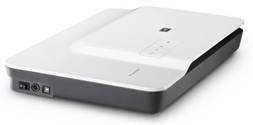
Fig. 1. Hp G3110 Scanjet Scanner (9000x4800 dpi resolution).
2.1. Scanner Installation Procedure and Settings
The scanner software can be installed into the computer operating system using the installation cd or by downloading the software set up from the manufacturer’s web site. After the scanner has been installed, the scanner icon will appear on the desktop. To customize the scanner settings, click on the scanner icon and then click on scan picture. This will lead to the page that has "change settings of the picture" menu. When you click on change settings, properties icon will appear, on it you will see dialogue box with "advanced picture settings". Select it and more options on the properties will appear. Then, click on or select the followings: restore faded colour, apply adaptive lighting, sharpen, remove dust and scratches, change the output resolution to 4800, scan multiple photos to individual files (single selection for each photo found), and higher bit depths (slower scan time). After all these changes then ok it to finalize the customize settings. The purpose is to have a scanner setting that will give very high resolution
2.2. Scanning Procedure
The first step of the scanning process is to place the palm or sole on the scanner, close the scanner cover to the extent the hand or feet can accommodate and then click on scan. Allow the exposure light which can been seen from the scanner glass surface to run through until it shows the scan preview on the computer screen. Once the scanned image has appeared on the computer screen, do the following: a) crop the picture to reduce the extra space surrounding the picture of the palm. b) click on correct picture menu and do the followings: change "remove dust" and "scratches" from normal to high, change "sharpen" from medium to extreme and click on descreen; c) Adjust the colour of the scan by clicking on "adjust colour" menu then select "invert colour" or "invert grayscale colour"; d) click on resize output and change percentage scale from 100 to 10 to reduce the size of the picture. e) Change the output type from colour to grayscale. After all these changes, click on "finish".
Then the scanner will do the final scanning. This is the second step in the scanning process. Because the scanning process is a bit slow, after the automatic re-scanning, it is possible to switch the scanner cord to any other USB ports on the computer to make it faster. At this point, a new scan can begin, while the former one is still running until it finishes by itself. When it has finished scanning, the scanned picture will be saved in scan folder. It can be resaved using appropriate file name for easy identification.
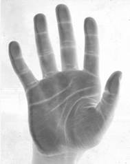
Fig. 2. Scanned Palm.
2.3. Analysis Procedure
Autocad software program is use to analyze the scanned image. The software can quantify image dimensions whether they are linear or angular. In dermatoglyphics, it can be use to count the ridges and also measure the angles with very high accuracy. The procedure involves opening the autocad work space by clicking on the icon in desktop. Paste the copied scanned palm or sole image on it. After pasting the picture, click on the zooming icon in "status bar" located below the page and drag on the picture to make it large enough for the triradius to be identified (a triradius is the meeting point of three ridges that form an angle. If after zooming, some parts of the picture are not showing then go to status bar and click on "pan". The pan is use in scrolling up and down in case where the scrolls are not there. Go to the "menu bar" and click on "dimension" and then on "line". This feature depends on the version of the autocad program, but it is in the 2010 version which we used as prototype. After clicking on line, use the cursor to draw a straight line from triradius "a" to "d" and from "d" to "t" and from "t" to "a" to form a triangle. Go again to the menu bar and click on dimension and then on "angular". Use the cursor and click on the lines and angular measurement will appear. For example, to measure "atd" angle, use the cursor and click on "a-t line" and "d-t line" and then angle measurement will pop up (Fig b). To get other angular dimensions, the same procedure is followed.
For finger ridge counting, a straight line is drawn from one triradius (i.e. triradial point) to the core (which is the middle of the pattern). For palmar ridge count, a straight line is also drawn from one triradius to the next triradius for example "a" triradius to "b" triradius. To execute this procedure, go to menu bar, click on "Draw", go to "point" and then select "multiple points". Go again and click on "format" select "point style" and then use any shape you want to use by clicking on it. Use the cursor and click on the ridges one after the other until the last ridge. Highlight all the points dotted and the number of the points will be shown on the left side of the computer which is the same as the number of the ridges counted (fig 3a)
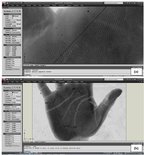
Fig. 3 a-b. Pictures showing the measurement of atd, dat and adt angles and the counting of the ridges using autocad program.
Table 1. Test for comparison of mean values for a-ridge count and atd angle for improvise digital and ink method.
| Parameter | Method | N | Mean +SE | Calculated t score | Critical t score at 0.05 level | Inference |
| a-b ridge count | Ink | 30 | 35.1 ±1.11 | 2.39 | 2.00 | p<0.05 |
| IDM | 30 | 38.1 ±0 .06 | ||||
| atd angle (degrees) | Ink | 30 | 44 ±1.35 | 2.10 | 2.00 | p<0.05 |
| IDM | 30 | 40±1.35 |
IDM = improvise new digital method
3. Results and Discussion
The accuracy of the method was tested by evaluating a-b ridge count and atd angle of 30 samples using the ink method and this new method as show in table 1. There was significant difference in the results (p<0.05). This difference was as a result of our ability to clearly identify the exact triradius point and the ridges which resulted in accurate measurement of angles and counting of ridges.
As already noted, the clarity of the prints of the hand and foot are very important in dermatoglyphic analysis. Poor print inevitably results in deficient data that would lead to wrong inference. Traditionally, the ink method as described by Cummins and Mild1 is still widely used in most developing nation like Nigeria. From experience, this had serious limitations especially when working with subjects with decrease mental and social interaction abilities such as Autistic and Down syndrome patients. These limitations include difficulty in the accurate application of ink on the hand and foot, in some cases leading to over inking. The inconvenience the ink posed to volunteers and the difficulty in erasing the ink from the hand. Similar observations were made by Gupta ad Gupta16 while comparing the lip stick method to the ink method. They reported an improved ease of obtaining the prints, clarity and accuracy of prints obtained, ease of analyzing prints and subjects friendliness of the procedure for lip stick method.
Lack of modern technological facilities is a serious limitation to quality scientific research in developing nations. In most cases, it is as a result of the cost of equipment and sometimes the technical knowledge. The field of dermatoglphics is not exempted from these limitations. Thus, this new method which makes use of easy-to-get facilities would give accurate results as the expensive customize biometric machine
An additional advantage of this method is that prints can be subjected to digital magnification. Digital magnification would afford the user the ability to clearly identify dermatoglyphic points and patterns that would have been obscured if ordinary hand lenses were used.
4. Conclusions
We therefore recommend this method because of easy availability of the materials needed for its operations. The scanner which is ordinarily use for the scanning of document in the office is quite cheap. Autocad program is readily available software which can be downloaded free from the internet. The method is easy to perform, not expensive as it involves only digital scanner and autocad software. It can be used in clinical, forensic and research settings in both rural and urban areas of developing countries where access to high tech biometric equipment is limited due to cost
References
- Cummins H. and Midlo C. (1943). Finger prints, Palm and soles. An introduction to dermatoglyphics Blakiston, Philadelphia.
- Reed T. and Meier R. (1990). How to take dermatoglyphic prints. Taking Dermatoglyphic print: A Self-instruction manual. Supplement to the Newsletter of the American Dermatoglyphics Association, 1-45.
- Pratibha R., Abhilash P.R., Herald J.S., Anuja N., Priya P. Chandrasekar T., Sentamilselvi G., Janaki V.R. (2011). Conventional Dermatoglyphics – Revived Concept: A Review. International Journal of Pharmacology and Biological Sciences, 2(3):446-457.
- Ozyurt B., Songur A., Sarislmaz M., Akyol O., Namli M., Demorel R. (2010). Dermatoglyphics as markers of prenatal disturbances in Schizophrenia: a case-control study Turk. Journal of Medical Sciences, 40(6):917-924.
- Boroffice R.A. (1978). Down’s Syndrome in Nigeria: dermatoglyphic analysis of 50 cases. Nigeria Medical Journal, 8(6):571-6.
- Arrieta M.I., Martinez B., Criado B., Simon A., Salazar L., Lostao C.M. (1990).Dermatoglyphic analysis of autistic Basque children. American Journal of Medical Genetic, 35(1):1-9.
- Milicic J., Bujas P.Z., Bozikov J. (2003). Dermatoglyphs of digito-palmar complex in autistic disorder: family analysis. Croatia Medical Journal, 44(4):469-76.
- Oladipo G.S., Okoh P.D., Oghenemavwe L.E., Yorkum L.K. (2013). Dermatoglyphic Patterns of Autistic Children in Nigeria. Journal of Biology, Agriculture and Healthcare, 3(7):80-83.
- Stosljevic M. and Adamovic M. (2013). Dermatoglyphic characteristics of digito-palmar complex in autistic boys in Serbia. Vojnosanit Pregl, 70(4):386-390.
- Rosa A., Gutierrez B., Guerra A., Arisa B., Fananas L. (2001).Dermatoglyphics and abnormal palmar flexion creases as markers of early prenatal stress in children with Idiopathic Intellectual disability. Journal of Intellectual Disability Research, 45(5):416-23.
- Seema M.A., Gandhi D., Singh M. (2012).Dermatoglyphics – Study and Review of Literature. Novel Science International Journal of Medical Science, 1(6):191-198.
- Gyenis G. (2000). A Short history and some results of the dermatoglyphic studies in Hungary, Acta Biologica Szegediensis, 44 (1-4):135-138.
- Sharma M.K., Jhawar P., Sharma H., Sharma S., Kalavatia I. (2012). Dermatoglyphics an attempt to predict Downs syndrome. International Journal of Biological & Medical Research, 3(2):1631-1635.
- Sharma M.K. and Sharma H. (2012). Dermatoglyphics: A Diagnostic Tool to Predict Diabetes. Journal of Clinical and Diagnostics Research, 6(3):327-332.
- Osunwoke E.A., Ordu K.S., Hart J., Esomonu C., Tamunokuro F.B. (2008). A Study on the Dermatoglyphic Patterns of Okrika and Ikwerre Ethnic Groups of Nigeria. Scientia Africana, 7(2):143-147.
- Gupta R.K. and Gupta A.K. (2013). New, Easy and Effective Method to take Dermatoglyphic Prints. National Journal of Medical Research, 3(1):45-47.
- Macarthur J.W., Ford N. (1937). A Biological study of the dionne quintuplets all Identical Set. Univ. Toronto Studies, Child Development Series, Univ. Toronto Press. 11: 49.
- NEC (2015). LiveScan Overview. http://ad.nec.com/en_All/solution/security-and-public-safety/biometrics/livescan-overview.html. Retrieved 15/03/2015.


